Event Information
Location: New York Academy of Medicine, 1216 Fifth Avenue at 103rd St
Click here for the schedule of events
About Us
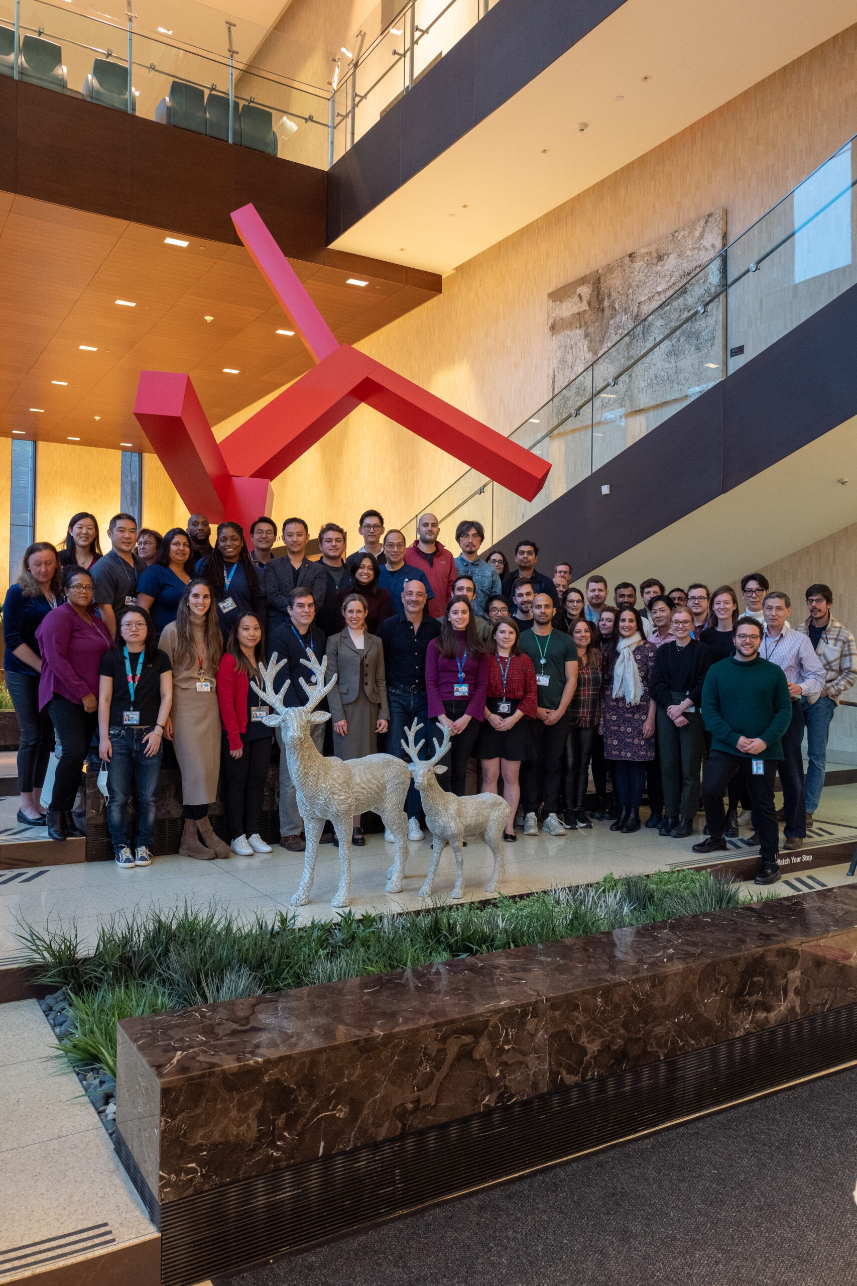
The BioMedical Engineering and Imaging Institute (BMEII) at the Icahn School of Medicine at Mount Sinai focuses on the use of multimodality imaging for brain, heart, and cancer research, along with research in nanomedicine for precision imaging and drug delivery. Our team conducts cutting edge research and serves as a research catalyst for a new generation of translational and molecular imaging methodologies to advance human health.
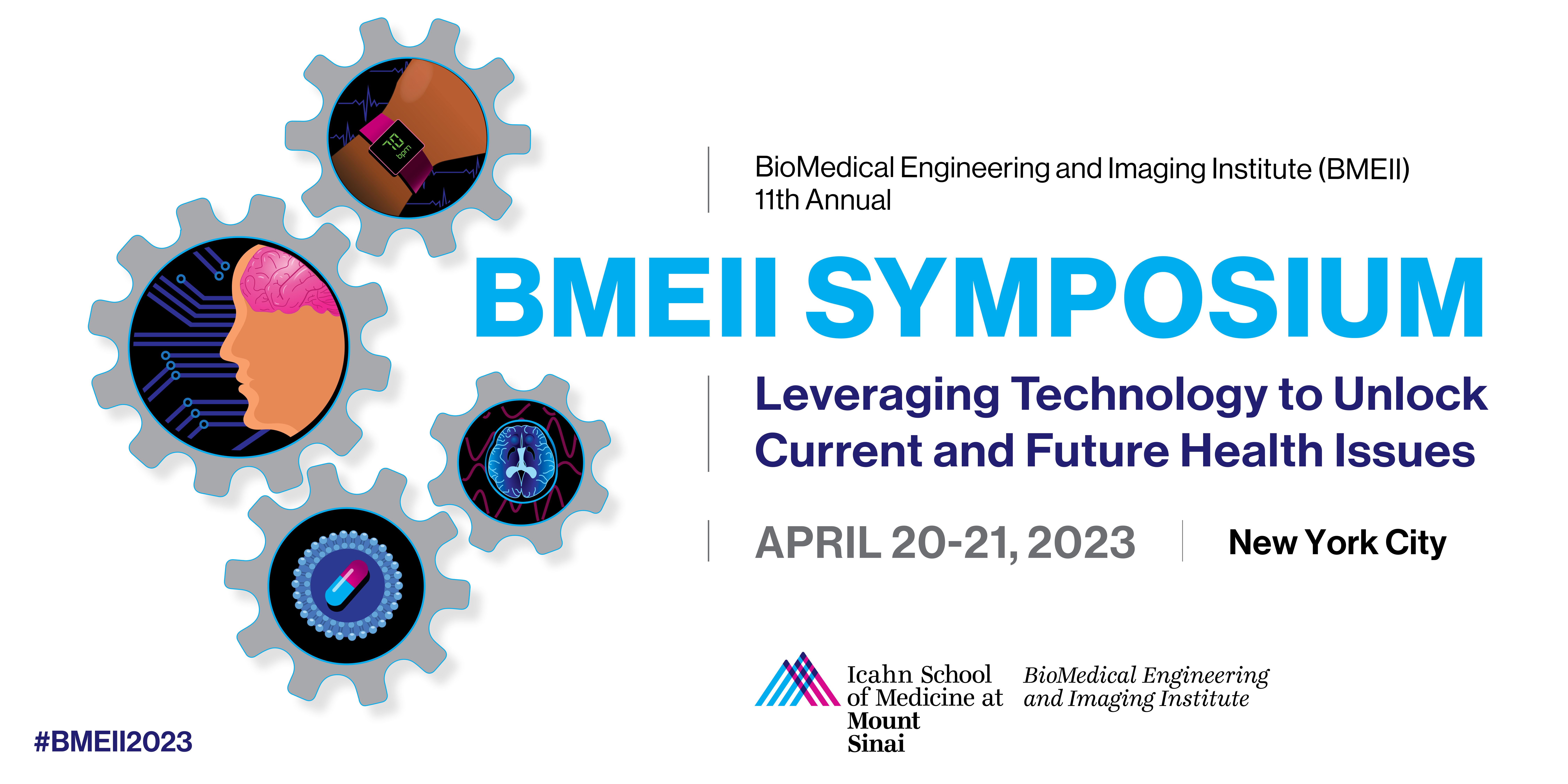
Now in its 11th year, the BMEII Symposium highlights the multidisciplinary nature of our institute and facilitates exchanges between innovators across the globe in the areas of biomedical imaging, nanomedicine, artificial intelligence, and wearable technology.
Registration
Please register below or visit our Eventbrite page here.
For sponsorship opportunities, please contact [email protected].
Call for Abstracts
In order to submit an abstract, you must register for the event. You will need to provide the 10 digit code of your ticket. Please use the provided abstract and submission form below. This year will, abstracts will be considered for oral or poster presentation as well as Innovation Station.
Deadline: March 8, 5pm ET
What is Innovation Station?
The Speakers

Michelle S. Bradbury, MD PhD
Memorial Sloan Kettering Cancer Center, NYC
Dr. Bradbury earned a B.A. in Chemistry from the University of Pennsylvania, an M.S. degree in Nuclear Engineering from the University of Maryland, and a PhD in Nuclear Engineering (Radiological Sciences) from the Massachusetts Institute of Technology. She received her MD degree at George Washington University School of Medicine, followed by an Internship in Surgery, Residency in Diagnostic Radiology, and Fellowships in both Neuroradiology at the Bowman Gray School of Medicine and Molecular Imaging at MSKCC. She currently holds appointments as a Professor of Radiology at MSKCC and Weill Cornell Medical College, as well as holds a joint appointment in the Molecular Pharmacology program at Sloan Kettering Institute. She has been a Co-Director of the MSKCC-Cornell Center for Translation of Cancer Nanomedicines, Director of Intraoperative Imaging, and Co-Chair of the Innovations and Technology Team at MSK. She co-developed the C dot technology, an ultrasmall core-shell silica nanoparticle translated to the clinic for a range of diagnostic and therapeutic applications. Under the auspices of a U54 Center of Cancer Nanotechnology Excellence grant awarded in 2015, she and her long-standing collaborator, Professor Ulrich Wiesner at Cornell University, made significant advances in the design, characterization, development, and clinical translation of ultrasmall inorganic probes for targeted cancer detection and treatment. Dr. Bradbury has previously served as a Principal Investigator (or co- investigator) of both early and advanced stage surgical trials that utilize the C dot platform for fluorescence-guided surgery, multimodal imaging, and/or drug delivery. She has more than 180 issued and pending patents worldwide related to the technology and its applications. Her patents have been licensed to Elucida Oncology, Inc, a company which she co-founded and currently serves on its Scientific Advisory Board member.
Nanshu Lu, PhD
University of Texas at Austin
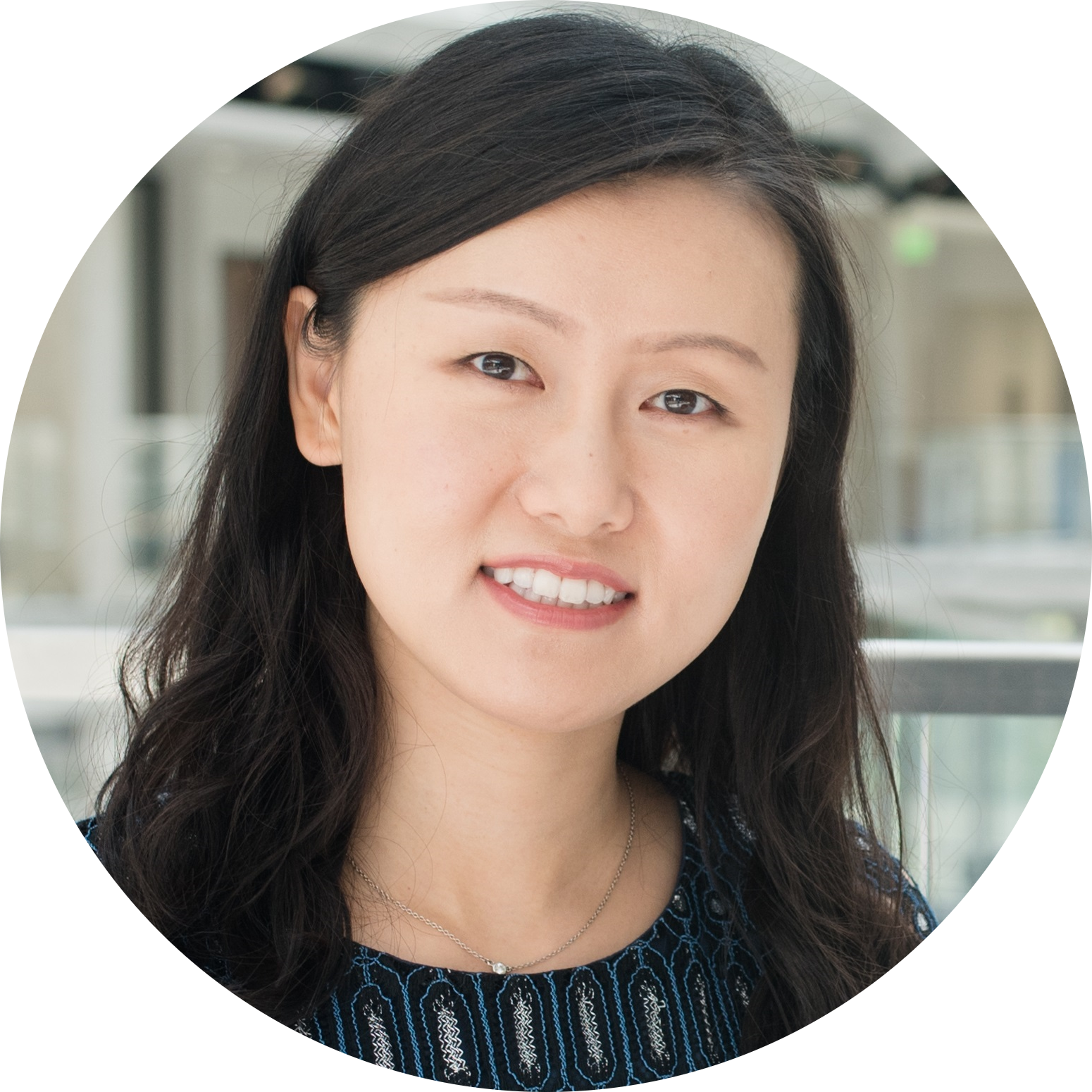
Dr. Nanshu Lu is the Frank and Kay Reese Professor at the University of Texas at Austin (UT). She received her B.Eng. with honors from Tsinghua University, Beijing, Ph.D. from Harvard University, and also Beckman Postdoctoral Fellowship at UIUC. Her research concerns the mechanics, materials, manufacture, and human / robot integration of soft electronics. She is an ASME Fellow and a Clarivate (Web of Science) highly cited researcher. She is currently an Associate Editor of Nano Letters and Journal of Applied Mechanics. She has been named MIT TR 35 and iCANX/ACS Nano Rising Star. She has received US NSF CAREER Award, US ONR and AFOSR Young Investigator Awards, 3M non-tenured faculty award, and the ASME 2022 Thomas J.R. Hughes Young Investigator Award. She has been selected as one of the five great innovators on campus and five world-changing women at UT. For more information, please visit Dr. Lu’s research group webpage at https://sites.utexas.edu/nanshulu/ and follow her on Twitter: @nanshulu.
Claudia Prieto, PhD
King’s College London, United Kingdom
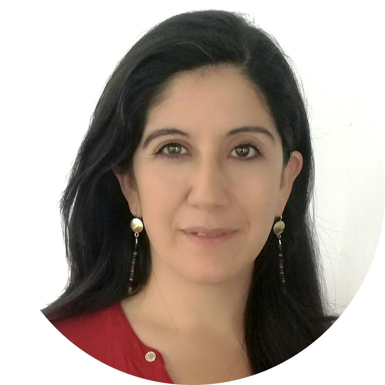
Claudia Prieto is a researcher known for her work in the fields of Magnetic Resonance Imaging (MRI), Artificial Intelligence (AI), and Computer Vision. Her main research concerns include Undersampling, Image Quality, AI, Computer Vision and Compressed Sensing. She is best known for her work in Motion Corrected Compressed Sensing for Free-Breathing Dynamic Cardiac MRI, Whole-Heart Coronary MR Angiography with 2D Self-Navigated Image Reconstruction, and Artificial Intelligence-Based Cardiac MRI Reconstruction. Throughout her career, she has published primarily in the fields of Artificial Intelligence, Computer Vision, and Image Quality. In recent years, her research has focused on the themes of Magnetic Resonance Imaging, Artificial Intelligence, Imaging Phantom, T2 Mapping, Biomedical Engineering, and Internal Medicine.
Evis Sala, MD, PhD
Università Cattolica del Sacro Cuore & Fondazione Policlinico Universitario Agostino Gemelli IRCC, Rome, Italy
Evis Sala is a Professor of Radiology and Chair of Department of Radiology at Università Cattolica del Sacro Cuore and Fondazione Policlinico Universitario Agostino Gemelli IRCC, Rome, Italy. Previously she was the Professor of Oncological Imaging at the University of Cambridge, UK and co-led the Cancer Research UK Cambridge Centre Advanced Cancer Imaging and the Integrated Cancer Medicine Programs. From 2012 to 2018, she served as the Professor of Radiology at Weil Cornell Medical College and Chief of Body Imaging Service at Memorial Sloan Kettering Cancer Center in New York.
Dr Sala’s research focuses on the development and validation of functional imaging biomarkers to rapidly evaluate treatment response using physiologic and metabolic tumour habitat imaging. Her research in the field of radiogenomics integrates quantitative imaging methods for evaluation of spatial and temporal tumour heterogeneity with genomics, proteomics and metabolomics. She is leading multiple research projects focusing on the development and implementation of artificial intelligence methods for image reconstruction, segmentation, and data integration.
Dr Sala is an outstanding educator, orator and mentor. In recognition for her contribution to education and research in oncological imaging she received the Radiology Society of North America (RSNA) Honoured Educator Award in 2014, 2017 and 2020. Her leadership extends to the most important international bodies in the field, as Honorary Member of RSNA, Fellow of the International Cancer Imaging Society, Fellow of the International Society for Magnetic Resonance in Medicine, Fellow of the European Society of Urogenital Radiology and Fellow of the Royal College of Physicians.

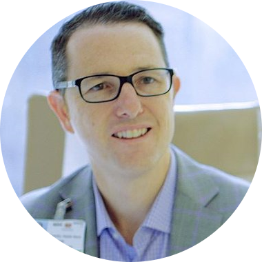
Brennan Spiegel, MD, MSHS
Cedars-Sinai Medical Center, California
Dr. Brennan Spiegel is director of health services research for Cedars-Sinai, where he directs a multidisciplinary team that investigates how digital health technologies, including wearable biosensors, smartphone applications, and virtual reality can improve people’s lives. His team developed one of largest and most widely documented healthcare VR program at Cedars-Sinai and helped to establish a new FDA-recognized field called medical extended reality, or “MXR” for short. Dr. Spiegel has published numerous books for medical and lay audiences and authored over 260 peer-reviewed articles. His digital health research has been featured by major media outlets, including the New York Times, Washington Post, and Wall Street Journal, among many others. Dr. Spiegel’s research is funded by the NIH, PCORI, and the California Initiative to Advance Precision Medicine. His virtual reality research won the 2017 “Webby Award” for best technology on the internet, and in 2020 he published the book VRx: How Immersive Therapeutics Will Transform Medicine, which was named by Wired Magazine as one of the top 8 science books of 2020.
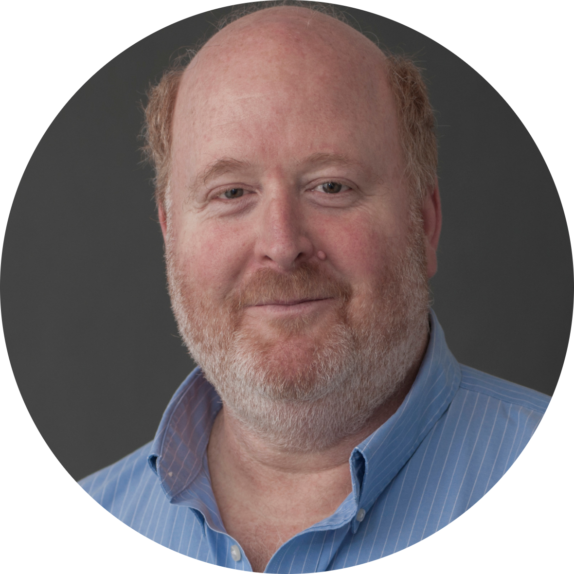
Lawrence Wald, PhD
Athinoula A. Martinos Center, Boston
Dr. Wald is a renowned expert in the field of high-field imaging of the brain. He has made significant contributions to the development of advanced imaging techniques, including the design and development of a 7 Tesla scanner and specialized coils for imaging human brain function. He has also been instrumental in the development of highly parallel phased array coils for use in both 3T and 7T scanners, and has made pioneering contributions to parallel transmit methods for B1+ mitigation in the head at 7T. Additionally, Dr. Wald has made important strides in the field of highly accelerated echo volume imaging, which has led to the development of new and more efficient imaging techniques. His work has been widely recognized and cited in the scientific community and has helped to push the boundaries of what is possible in the field of high-field imaging of the brain.
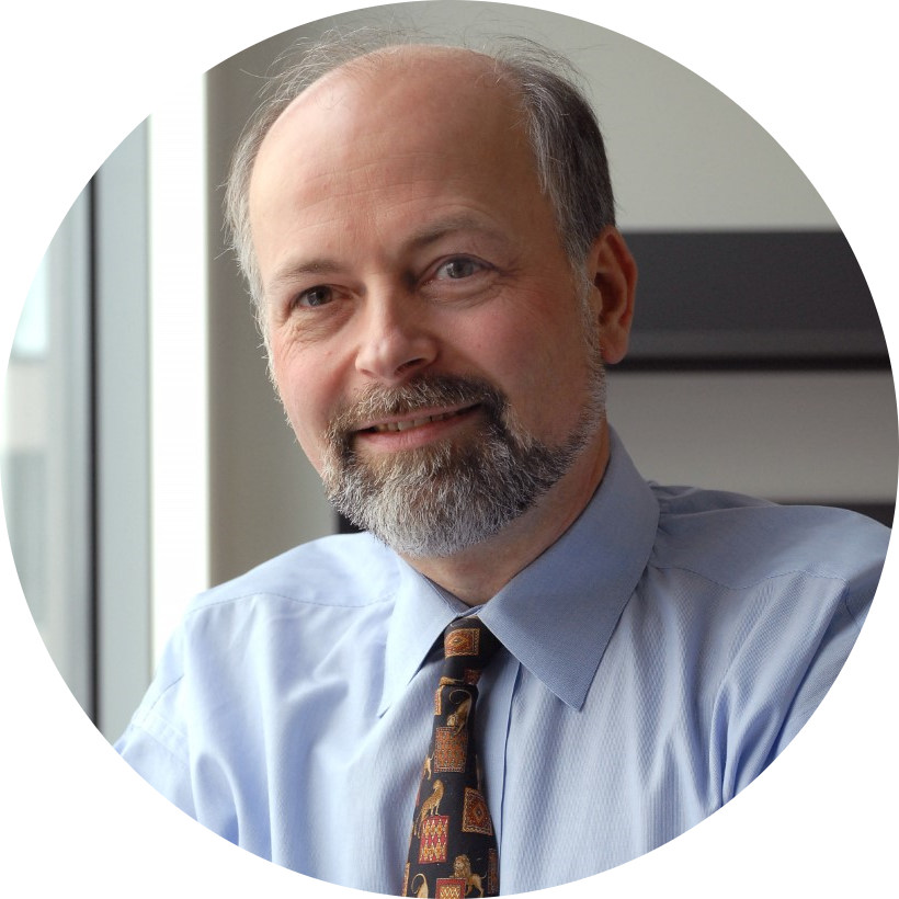
Ralph Weissleder, MD, PhD
Harvard University
Dr. Weissleder is the Thrall Professor Professor of Radiology at Harvard Medical School, Director of the Center for Systems Biology at Massachusetts General Hospital (MGH), and Attending Clinician (Interventional Radiology) at MGH. Dr. Weissleder is also Professor of System Biology and a faculty a member of the Department of Systems Biology at HMS and the Dana Farber Harvard Cancer Center. The focus of his research lab is to obtain a deeper understanding of human biology in health and disease, to translate new biological understanding into clinically useful diagnostics and to identify new therapeutic approaches and drug targets. His research has been translational and several of his developments have been licensed to companies and led to advanced clinical trials. He has published nearly 1,000 publications in peer reviewed journals (h-index 212) and has authored several textbooks. He is a member of the National Academy of Medicine, the American Academy of Arts and Sciences, the National Academy of Inventors and the German Academy of Sciences (Leopoldina). Website: csb.mgh.harvard.edu
Program
Day 1: April 20, 2023
2:00pm Registration – Day 1
2:30pm Opening Remarks
Zahi Fayad, PhD | Director, BMEII
Neil Rofsky, MD, MHA | Chair, Diagnostic, Molecular and Interventional Radiology
3:00pm Keynote: Ralph Weissleder, MD, PhD | Harvard Medical School
Leveraging Technology in Medicine
4:00pm Innovation Station / Poster Session / Welcome Reception
6:00pm Event Ends
Day 2: April 21, 2023
8:00am Breakfast and Registration – Day 2
8:30am Opening Remarks:
Zahi Fayad, PhD | Director, BMEII
Dennis Charney, MD | Anne and Joel Ehrenkranz Dean, Icahn School of Medicine at Mount Sinai
9:00am Session 1: Nanomedicine, Drug Delivery, and Cancer
Speaker 1: Michelle Bradbury, MD, PhD | Memorial Sloan Kettering Cancer Center
Engineered Ultrasmall Particle Treatment Tools for Clinical Cancer Care
Speaker 2: Evis Sala, MD, PhD | Università Cattolica del Sacro Cuore
Building Clinical Value in Cancer Care with MRI
10:45am Break
11:00am Oral Presentations
12:00pm Lunch
12:45pm Trivia
1:30pm Session 2: Advances in Medical Imaging
Speaker 1: Larry Wald, PhD | Harvard Medical School
New scanner architectures for brain imaging: portable/accessible low-field MRI and a human magnetic particle imager for sensitive functional imaging
Speaker 2: Claudia Prieto, PhD | Kings College London
Multiparametric 3D Cardiovascular MRI: All-in-one
3:15pm Break
3:30pm Session 3: Wearable Technologies and Virtual Reality Therapy
Speaker 1: Nanshu Lu, PhD | University of Texas at Austin
Wireless E-Tattoos for Ambulatory Multimodal Biometric Sensing
Speaker 2: Brennan Spiegel, MD, MSHS | Cedars-Sinai Medical Center
Virtually Better: The Art and Science of Medical Extended Reality
5:15pm Closing Remarks & Presentation of Awards
Neil Rofsky, MD, MHA
6:00pm Art Show & Reception
8:00pm Event Ends
Call for Artwork

The closing event of the BMEII Symposium, Windows to Our Body, is an art exhibition featuring scientific art drawn from the creative expressions of scientists and trainees. The art show will be printed and displayed for an auction to raise money for charity. All participants are encouraged to submit their work.
Deadline: March 8th, 2023 5 pm ET
News
[VIDEO] Aspen Ideas Festival: Dr. Fuchs and Dr. Fayad on Powering Medicine with Technology and Data Science
Zahi A. Fayad, PhD, Director of Biomedical Engineering and Imaging Institute sits down with Thomas Fuchs, DrSc, Dean of Artificial Intelligence and Human Health to discuss powering medicine with technology and data science. Click the image to watch the video.
[VIDEO] Dr. Zahi Fayad: Powering Medicine with Technology and Data Science
BMEII Director Zahi Fayad discusses the role of data science in medicine at the 2022 Aspen Ideas Festival. Click the image to watch the video.