Engineering and Medicine Seminars Series
Join us for our seminars, where we ask prominent researchers in their fields to share their work with us.

Previous Seminars


AEIT Seminars 2025-2026
Below you can see the list of the past BMEII Seminars, which were part of the AIET Seminar during the academic year 2025-2026.
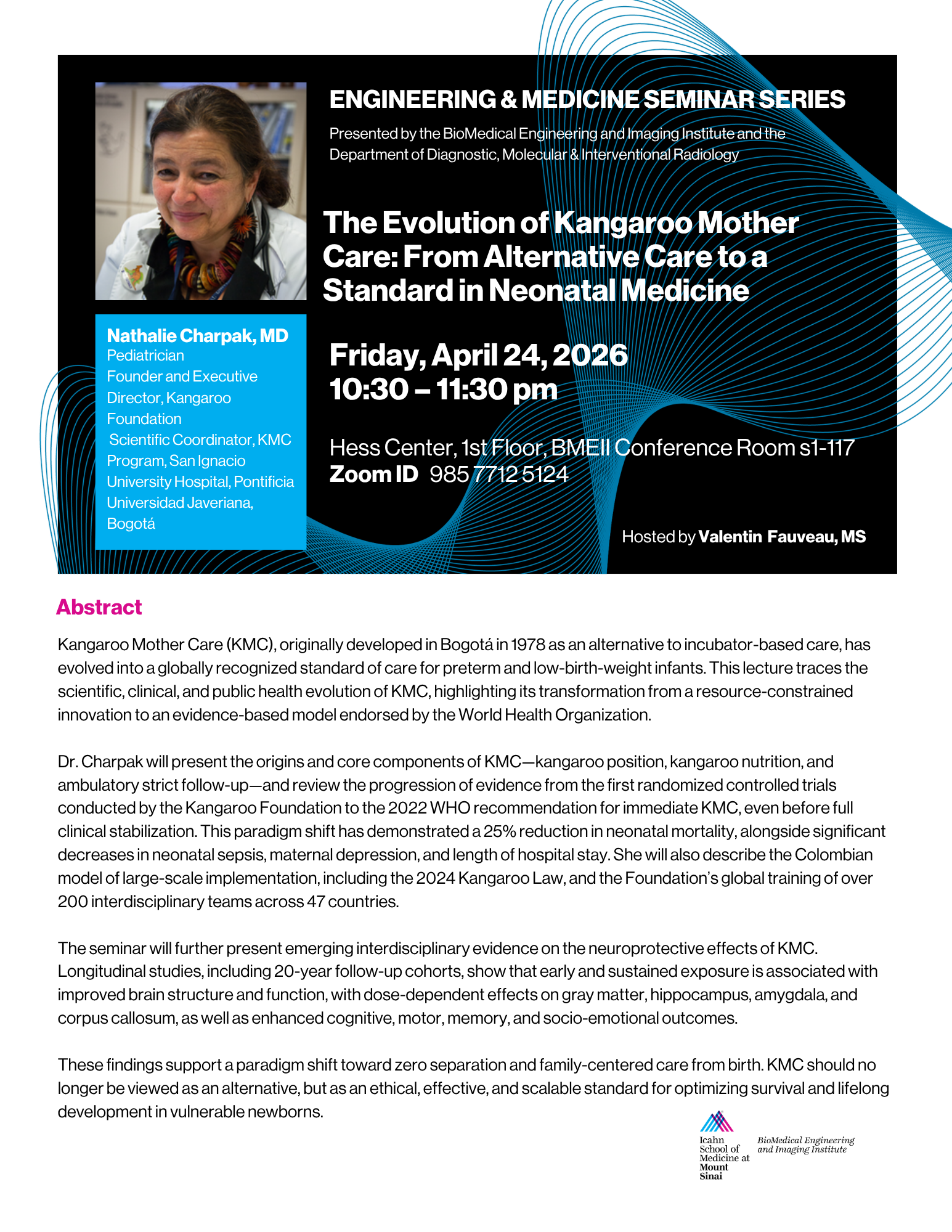
"The Evolution of Kangaroo Mother Care: From Alternative Care to a Standard in Neonatal Medicine" by Nathalie Charpak, MD.
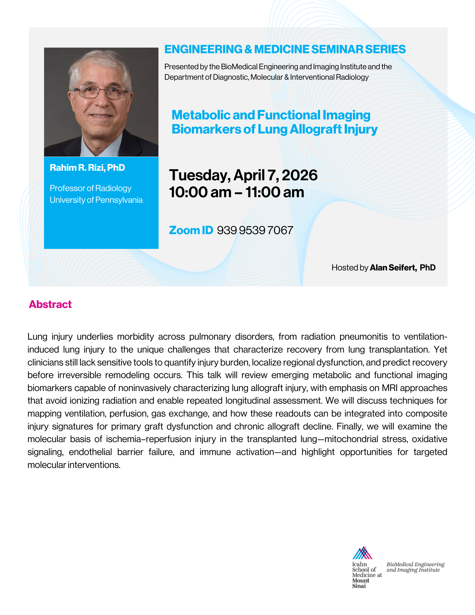
"Metabolic and Functional Imaging Biomarkers of Lung Allograft Injury" by Rahim R. Rizi, PhD
Abstract
Lung injury underlies morbidity across pulmonary disorders, from radiation pneumonitis to ventilation‑induced lung injury to the unique challenges that characterize recovery from lung transplantation. Yet clinicians still lack sensitive tools to quantify injury burden, localize regional dysfunction, and predict recovery before irreversible remodeling occurs. This talk will review emerging metabolic and functional imaging biomarkers capable of noninvasively characterizing lung allograft injury, with emphasis on MRI approaches that avoid ionizing radiation and enable repeated longitudinal assessment. We will discuss techniques for mapping ventilation, perfusion, gas exchange, and how these readouts can be integrated into composite injury signatures for primary graft dysfunction and chronic allograft decline. Finally, we will examine the molecular basis of ischemia–reperfusion injury in the transplanted lung—mitochondrial stress, oxidative signaling, endothelial barrier failure, and immune activation—and highlight opportunities for targeted molecular interventions.
Bio
Dr. Rizi has been performing researchon the non-invasive assessment of lung function, metabolism, and structure for the last three decades. The primary mission of his laboratory, the Functional and Metabolic Imaging Group (FMIG), is the development and application of novel hyperpolarized gas and liquid MRI techniques for examining pulmonary disorders. By drastically increasing the population difference between nuclear spin states, hyperpolarization provides a platform for imaging lung physiology and metabolism with spatial and temporal resolution unattainable with conventional MRI. FMIG continues to develop new imaging techniques with an eye toward several ultimate goals: the identification of changes in pulmonary function, metabolism, and structure associated with disease; a more complete understanding of pathogenesis; the establishment of a more sensitive testing environment to develop novel treatments for lung disease; and the evaluation of lung graft viability before transplant. Dr. Rizi’s significant contributions to the field of hyperpolarized gas MRI include the development of new imaging techniques for the comprehensive description of lung physiology and structure through the accurate measurement of parameters such as regional ventilation and alveolar oxygen tension, as well as the preclinical and clinical application of these techniques to study various pulmonary disorders.
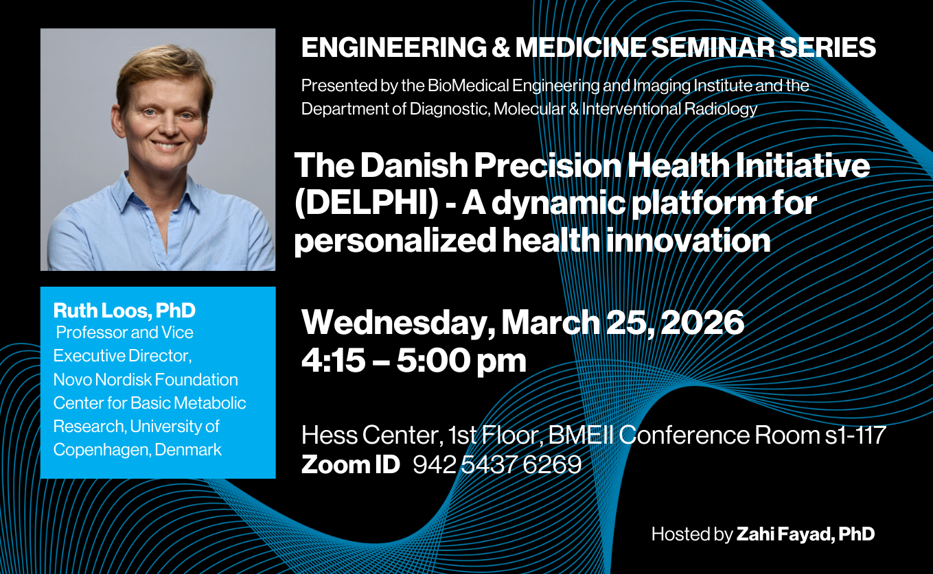
"The Danish Precision Health Initiative (DELPHI) - A dynamic platform for personalized health innovation" by Ruth Loos, PhD
Bio
Ruth Loos, PhD is a leading researcher in the genetics of obesity and metabolic health, currently serving as Vice Executive Director and Professor at the Novo Nordisk Foundation Center for Basic Metabolic Research at the University of Copenhagen. Her work focuses on identifying genetic and environmental determinants of body weight regulation, contributing to major gene-discovery efforts through the GIANT consortium. She also advances precision health approaches by integrating genetic, clinical, and lifestyle data to improve prediction, prevention, and treatment of obesity. Previously, she held multiple leadership roles at the Icahn School of Medicine at Mount Sinai, where she directed research on obesity and metabolic traits.
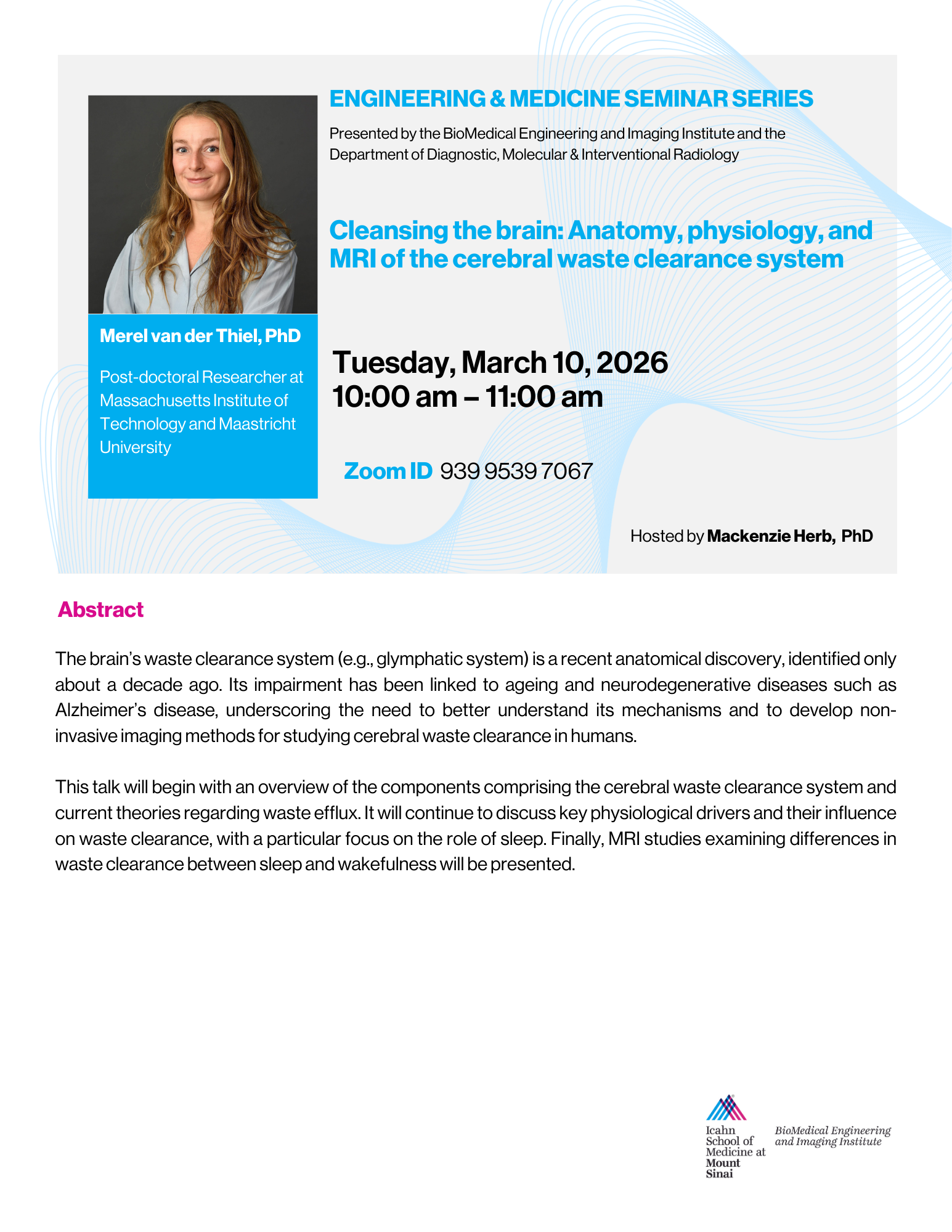
"Cleansing the brain: Anatomy, physiology, and MRI of the cerebral waste clearance system" by Merel van der Thiel, PhD
Abstract
The brain’s waste clearance system (e.g., glymphatic system) is a recent anatomical discovery, identified only about a decade ago. Its impairment has been linked to ageing and neurodegenerative diseases such as Alzheimer’s disease, underscoring the need to better understand its mechanisms and to develop non-invasive imaging methods for studying cerebral waste clearance in humans.
This talk will begin with an overview of the components comprising the cerebral waste clearance system and current theories regarding waste efflux. It will continue to discuss key physiological drivers and their influence on waste clearance, with a particular focus on the role of sleep. Finally, MRI studies examining differences in waste clearance between sleep and wakefulness will be presented.
Bio
Dr. Merel van der Thiel developed a strong interest in neurodegenerative diseases during her bachelor’s degree in Psychology (Cognitive Neuroscience track) at Leiden University, the Netherlands. This interest led her to pursue two MSc degrees with distinction: first in Neuroscience in Geneva, Switzerland, followed by a specialized MSc program in Dementia at University College London (UCL).
She subsequently obtained her PhD at Maastricht University, the Netherlands, where she specialized in advanced MRI methods to quantify components of the cerebral waste clearance system in relation to Alzheimer’s disease. She was awarded an Early Career Grant from the Dutch Alzheimer Society to continue her postdoctoral research at Maastricht University, focusing on autonomic influences on cerebral waste clearance.
Dr. van der Thiel is currently a visiting researcher at the Massachusetts Institute of Technology (MIT), USA, in the laboratory of Dr. Laura Lewis, where she investigates interstitial fluid movement using diffusion MRI during wakefulness and sleep.
"Towards Intelligent Wearable Assistants for Everyday Physical Interaction" by Yiyue Luo, PhD
Abstract
Everyday life is shaped by continuous physical interactions, touching, grasping, and moving, that underlie human behavior, health, and well-being. Intelligent wearable assistants aim to capture and enhance these interactions by seamlessly sensing, modeling, and responding to the body’s physical dynamics, providing clinicians and patients with actionable insights that were previously inaccessible. In this talk, I will present a series of textile-based systems that move beyond passive sensing to demonstrate the potential of intelligent wearables as everyday assistants. Examples include machine-knitted tactile garments that sense and guide motion, adaptive embroidered gloves for interactive tasks, and intelligent carpets that monitor whole-body activities. Together, these prototypes illustrate how textiles can serve as scalable and versatile platforms for building intelligent assistants that integrate into daily routines, bridging human physical interactions with computational intelligence. Together, these prototypes highlight how textiles, an everyday material, can become a powerful platform for intelligent health technologies, opening new pathways for prevention, early detection, and personalized rehabilitation.
Bio
Yiyue Luo is an Assistant Professor at University of Washington ECE. She received my Ph.D. degree in EECS from MIT in 2024, advised by Professor Wojciech Matusik and Professor Tomas Palacios. Before joining MIT, she received my B.S. in Materials Science and Engineering at University of Illinois Urbana-Champaign, working with Professor John A. Roger. Her research lies at the intersection of digital fabrication, human-computer/robot interaction, and applied AI. Her research on integrated intelligent textiles has been published in interdisciplinary journals, e.g., Nature Electronics and Nature Communications, top human-computer interactions, robotics and learning venues, e.g., CHI, UIST, CVPR, ICRA. Her work has been selected as the cover of Nature Electronics, awarded Best Paper at CHI, Best Paper Honorable Mention at UIST, featured in prominent media outlets, and invited to public museums and world congress exhibitions. She has been supported by fellowship from MathWorks, Google, and Accenture, and was recently listed as 30 under 30 North American 2024. She has been awarded funding from the UW Royal Research Fund and the CoMotion Entrepreneurship Grant.
"CEST MRI for Molecular Brain Imaging and AI-Driven Analysis" by Jianpan Huang, PhD
Abstract
Chemical exchange saturation transfer (CEST) magnetic resonance imaging (MRI) is a non‑invasive molecular imaging technique that enables in vivo detection of molecules containing exchangeable protons. It has been applied to visualize molecular changes in a range of neurological disorders, including brain tumors, Alzheimer’s disease, and multiple sclerosis. Over the past decade, CEST MRI has gained traction in numerous preclinical and clinical imaging centers worldwide. Despite its promising potential, widespread clinical translation of CEST MRI is hindered by challenges such as complex quantification methods and limited spatial resolution. Artificial intelligence (AI) in medical imaging is a rapidly evolving field that leverages neural networks to enhance, analyze, and interpret complex datasets. This talk will introduce the basic principles of CEST MRI and review its applications in imaging neurological diseases. It will also highlight recent advances in applying AI to CEST MRI, including techniques for streamlining molecular quantification and improving image quality. AI-assisted CEST MRI has the potential to improve diagnostic accuracy and support more informed clinical decision‑making.
Bio
Dr. Jianpan Huang is an assistant professor in the Department of Diagnostic Radiology and deputy director of the Tam Wing Fan Neuroimaging Research Laboratory at the Li Ka Shing Faculty of Medicine, The University of Hong Kong. He obtained his Bachelor’s and Master’s degrees from Xiamen University and Ph.D. from City University of Hong Kong. In 2022, Dr. Huang was named a Junior Fellow of the International Society for Magnetic Resonance in Medicine (ISMRM). He also received the Young Investigator Award from the Overseas Chinese Society for Magnetic Resonance in Medicine (OCSMRM) in 2020 and from the International Chemical Exchange Saturation Transfer (CEST) Workshop in 2018.
Dr. Huang has published around 50 peer-reviewed jounal papers in leading international journals, including Science Advances, Nature Communications, JMRI, MRM, and IEEE JBHI, and holds nine granted patents. He serves as a reviewer for over 20 journals, including JMRI, MRM, European Radiology and Comunications Physics, and also a grant reviewer for UK Research and Innovation and the Israel Science Foundation. His current research focuses on magnetic resonance imaging (MRI), chemical exchange saturation transfer (CEST) MRI, artificial intelligence (AI), and neuroscience.
"Multicentre Standardisation of Renal MRI" by Sue Francis, PhD
Abstract
This talk will outline developments on multicentre renal MRI data acquisition and analysis showing examples from the UKRIN-MAPS (MRI Acquisition and Processing Standardisation) project which aimed to develop harmonized renalMR protocols across vendors. Results of the repeatability of the UKRINMAPS multi-parametric protocol will be shown using a “travelling kidney” study, scanning healthy subjects across multiple sites. Analysis pipelines for automated analysis including AI segmentation methods will be shown. The talk will demonstrate the application of this x-vendor protocol to the AFIRM study which has collected renal MRI data in Chronic Kidney Disease patients at baseline and 2 years.
Bio
Prof Sue Francis is a physicist with 25 years’ experience on developing MRI methods for biomedical applications and translation. She works across the brain and body and across MR field strengths. Since 2005, she has led a programme of work exploiting the capabilities of functional and anatomical high and ultra-high field MRI in neuroscience, and is currently involved with the development of the 11.7T human National Facility in Nottingham which is due to be at field in 2027. In 2009, she developed a programme of imaging in quantitative imaging of the body, and leads work at Nottingham on the development of MRI methods to study renal and liver function. She initiated the UK Renal Imaging Network (UKRIN) and led the UKRIN-MAPS MRC Partnership grant to develop a harmonized approach in renal MRI across MR vendors. Her research interests include the development and application of multiparametric quantitative MRI methods, with a particular interest in Arterial Spin Labelling (ASL) methods to non-invasively assess tissue perfusion in the brain, kidney and liver. She has published widely on the application of renal MRI to study physiological modulations and disease pathophysiology.
"Profiling liver pathway through biomechanics, Quantifying response to therapy, and “Seeing” vascular architecture" by Ralph Sinkus, PhD
Abstract
Magnetic Resonance Elastography (MRE) has emerged as a powerful non-invasive modality to quantify tissue biomechanics, extending the clinical intuition of manual palpation into a quantitative, spatially resolved imaging biomarker. In this work, we present recent advances in gravitational transducer–based 3D MRE and demonstrate how full complex shear modulus measurements enable refined characterization of liver disease pathways, assessment of therapeutic response in oncology, and inference of underlying vascular architecture.
A robust gravitational MRE (gMRE) driver, based on an eccentric rotating mass, was developed to deliver stable, harmonic-free mechanical excitation with high efficiency. Following its translation from an EU Horizon 2020 research project into an FDA-approved clinical product, the technology enables reproducible whole-organ 3D MRE acquisitions integrated into standard clinical MRI workflows. Unlike conventional 2D MRE, which reports only the magnitude of the complex shear modulus |G*|, 3D MRE provides access to both stiffness and wave attenuation (phase angle), yielding enhanced sensitivity to microstructural changes.
In the liver, we show that 3D MRE identifies a pre-fibrotic biomechanical niche characterized by reduced shear wave attenuation despite only modest increases in stiffness, consistent with early collagen III deposition from activated hepatic stellate cells. This biomechanical signature precedes conventional fibrosis markers and correlates with biochemical tests and histology, enabling early detection of liver damage and identification of mechanical states associated with increased hepatocellular carcinoma (HCC) risk. Beyond hepatology, we demonstrate the value of MRE for early response assessment to neoadjuvant chemotherapy in breast cancer, where collagen remodeling following initial treatment cycles predicts therapeutic outcome with higher precision than current imaging standards.
Finally, leveraging wave dispersion induced by multiple scattering, we illustrate how macroscopic MRE measurements can probe microscopic vascular architecture, bridging several orders of magnitude in scale and opening new avenues for non-invasive characterization of tumor microenvironments and treatment response. Together, these results position advanced 3D MRE as a versatile, physics-informed biomarker platform for precision medicine across liver disease and oncology.
Bio
I am a physicist with a background in high-energy and nuclear physics as well as MRI, leading a translational research team in Paris (University Paris Cité, Sorbonne Paris Cité, Bichat/Beaujon Hospitals). My career spans both academia and industry. During my PhD at DESY (Hamburg, Germany), I worked on quantum electrodynamics/chromodynamics and developed a neural-network system for electron identification in particle collisions. I then joined Philips Medical Systems, focusing on MRI and MR-elastography.
In 2004, I transitioned to academia, establishing an MRI group at ESPCI Paris and collaborating with Supersonic Imaging. I secured a permanent CNRS research director position in 2007 and developed a multidisciplinary program integrating work at molecular, animal, and clinical scales.
In 2013, I became Chair in Biomedical Engineering at King’s College London. With collaborators in modeling and oncology, we led a Horizon 2020 project to measure tumour forces as indicators of therapy response. This research led to a product developed by QED and adopted by Siemens.
Our teams in London and Paris now focus on quantifying tissue biomechanics across organs and scales—from liver, breast, and brain to neuronal and organoid systems. In Paris, we address critical questions in liver oncology: identifying precancerous niches, predicting therapy response, and developing imaging biomarkers such as tumour pressure and tissue stiffness. Our clinical partnerships enable direct hypothesis testing at the patient level.
"Actuator-Enhanced Imaging and Diagnostics for Personalized Healthcare and Physiological Modeling" by June Ueda, PhD
Abstract
This talk presents recent advancements in actuator-enhanced imaging and diagnostics for personalized physiological modeling, with a focus on Magnetic Resonance Elastography (MRE) and human motor system identification. Building on the concept of digital twins in healthcare, the research explores how mechanical perturbations—delivered via robotic and motion control platforms—can improve the accuracy of individualized models for both organ diagnostics and neuromuscular control. Emphasis will be placed on system identification techniques, including the generation of mechanical perturbations optimized for upper limb impedance estimation using spectral flatness criteria. The talk will also highlight innovations in actuator design, such as tunable-frequency piezoelectric systems for spinal disc imaging and MRI-compatible robotic dosimeters for implant safety assessment. Key applications include the development of automated quality control systems for MRE using deep learning to assess diagnostic image quality and enhance liver stiffness measurement. These technologies collectively advance the reliability, efficiency, and personalization of physiological diagnostics and therapeutic planning.
Bio
"Metabolic MRI: Emerging Technologies for Clinical Translation" by Daniel Paech, MD, PhD, MSc
Abstract
Metabolic imaging technologies have the potential to transform the field of diagnostic radiology by offering novel insights into tissue function and disease. These technologies, including advanced MR spectroscopy, chemical exchange saturation transfer (CEST) MRI, and X nuclei imaging, enable non-invasive characterization of metabolic processes in vivo. This presentation will discuss recent advances in metabolic MR imaging, their clinical applications in diagnosing and monitoring diseases, and the challenges in translating these methods from research to practice. Highlights will include applications in neuro-oncology and neurodegenerative diseases. The role of new MR technologies, such as ultra-high field strength and high-performance gradient systems, will also be highlighted for their potential to further enhance metabolic imaging capabilities.
Bio
Daniel Paech, MD, PhD, MSc, is an Associate Professor of Radiology at Harvard Medical School and Medical Director of the Brigham Research Imaging Core and the Mass General Brigham (MGB) Ultra-High-Field MRI Center. He is a board-certified radiologist and neuroradiologist (diagnostic and neuro-interventional) and serves as attending neuroradiologist in the Division of Neuroradiology at MGB.
Dr. Paech’s research focuses on the development and clinical translation of novel imaging technologies, with a particular emphasis on ultra-high-field MRI. His expertise is in metabolic imaging, with applications in neuro-oncology, neurodegenerative disease, and ischemic stroke.
Passcode: O9T01G@q
"The Case of the Hidden Signatures: Designing Imaging AI to Bridge Patterns, Predictions & Precision Medicine" by Satish E. Viswanath, PhD
Abstract
Developing artificial intelligence (AI) schemes to assist the clinician towards enabling precision medicine approaches requires development of objective markers that are predictive of disease response to treatment or prognostic of longer-term patient survival. The solutions being developed in my group in this regard involve designing computational imaging features together with histology or molecular data for detailed tissue and disease characterization in vivo as well be associated with patient outcomes. The key innovation in this approach lies in “handcrafting” unique tools that can capture biologically relevant and clinically intuitive measurements from routinely acquired imaging (MRI, CT, PET) or digitized images of tissue specimens. Further, by conducting cross-scale associations between imaging, pathology, and -omics, we can not only “unlock” and integrate the information captured by these different, disparate data modalities but also develop an interpretable and intuitive understanding of what drives their performance. Specific problems addressed via the new computerized imaging markers we have developed include: (a) predicting response to treatment to identify optimal therapeutic pathways, as well as (b) evaluating therapeutic response to guide follow-up procedures. We will further examine how to account for differences between sites, scanners, and acquisition parameters to ensure generalizable performance of AI tools and computational imaging features; crucial for wider clinical translation and widespread adoption. These will be discussed in the context of clinical applications in colorectal and renal cancers, digestive diseases, as well as pediatric conditions.
Bio
Dr. Viswanath is an Associate Professor in the Departments of Pediatrics and Biomedical Engineering at Emory University, since Fall 2025. He is also a Research Scientist & Biomedical Engineer at the Cleveland VA Medical Center. Previously, he was an Associate Professor of Biomedical Engineering at Case Western Reserve University. The primary focus of his research has been developing new artificial intelligence (AI) approaches including image analytics, radiomics, and machine learning schemes; applied to problems in computer-aided diagnosis & detection, disease characterization, as well as quantitative evaluation of response to treatment; in gastrointestinal cancers and digestive diseases. He has authored over 55 peer-reviewed journal publications, over 120 conference papers & abstracts, 1 book chapter, as well as delivered over 90 invited talks and panel discussions both in the US and abroad. He has 10 issued patents in the areas of medical image analysis, computer-aided diagnosis, and pattern recognition. Dr. Viswanath is an Associate Editor or Editorial Board Member for 9 leading international peer-reviewed journals, serves as Program Committee Member or Area Chair for 3 major medical imaging conferences, and has been elected to Senior Member in the National Academy of Inventors, the IEEE, and the SPIE. He has been selected for the Fulbright Specialist Award, in addition to multiple awards from SIIM, SPIE, and Crain’s Cleveland Business. His lab’s research has been funded since 2016 through the DOD/CDMRP, the NIH (NCI, NIBIB, NINR, NHLBI), the VA, and the State of Ohio.
Passcode: hbNGs6&8
"AI-powered Diagnosis and Prognosis of Musculoskeletal Diseases" by Cem M. Deniz, PhD
"AI-enabled precision medicine for cancer research and innovations in medical devices" by Gautham Pasupuleti
"Quantitative MRI of Adipose Oxygenation and Hypertrophy in Type 2 Diabetes" by Scott C Beeman, PhD
"PET and MRI for a Holistic View of Glymphatic Function" by Chuan Huang, PhD
Abstract
The glymphatic system is increasingly recognized as a critical pathway for brain waste clearance, with implications for aging and neurodegenerative diseases. However, imaging glymphatic function in humans remains a major challenge due to the system’s complex, multiscale dynamics. In this talk, I will present a multimodal imaging approach using PET and MRI to achieve a more comprehensive and clinically translatable assessment of glymphatic function.
I will review the limitations of current MRI-based methods—including phase-contrast MRI, DWI, and DTI-ALPS—which primarily infer fluid motion indirectly and often lack sensitivity to the slow and heterogeneous flow patterns characteristic of glymphatic transport. In contrast, PET imaging enables direct quantification of solute clearance over time, offering a complementary view of net mass transport. I will highlight recent work using dynamic 18F-FDG PET to quantify ventricular CSF clearance and demonstrate its sensitivity to age-related decline, test–retest reliability, and independence from brain metabolism.
Bio
Dr. Chuan Huang is an Associate Professor of Radiology and Imaging Sciences and the Director of PET-MRI Research at Emory University School of Medicine. He was named a Distinguished Investigator by the Academy for Radiology & Biomedical Imaging Research in 2023.
Dr. Huang’s research focuses on PET/MR neuroimaging and its broader clinical and translational applications. He has authored over 70 peer-reviewed publications and holds three patents. Prior to joining Emory in November 2022, he was a tenured Associate Professor of Radiology and Psychiatry at the State University of New York at Stony Brook. He completed his postdoctoral training at Massachusetts General Hospital and Harvard Medical School in 2014, where he was subsequently promoted to Instructor.
He previously served as Chair of the ISMRM PET/MR Study Group and was a member of the organizing committee for the 2023 SNMMI–ISMRM co-sponsored PET/MR Workshop. He currently serves on the committee of the ISMRM MR in Psychiatry Study Group.
Passcode: ^DZq9$*6
"The MRI RF Coil Array Data Hub: From Imaging to Satellite Connectivity" by Dean Darnell, PhD
Abstract
All MRI scanners must have an RF coil or array for transmitting/receiving the RF signal during imaging regardless of field strength or magnet design. In this talk, we will explore the next generation of multi-purpose integrated RF/wireless (iRFW) coil designs, which can perform simultaneous MR signal reception and wireless data transfer by allowing RF currents at the Larmor frequency and in a wireless communication band(s) to flow on the same conductor. These coils can wirelessly transmit MRI images, enable wireless MRI clock synchronization via Global Navigation Satellite System (GNSS) signals from satellite atomic clocks, and support wireless ultrasound-based respiratory monitoring from within the bore to the scanner console or cloud. In low-field applications, the iRFW coil design, termed an iRFW-Cellular coil design, can further enable wireless data transmission of image data over cellular or satellite networks while imaging. In total, the RF coil array is not only essential for imaging but a natural data hub for wirelessly moving data out of the scanner bore.
Bio
Dr. Darnell earned his PhD in physics in 2005 and spent eight years at Apple Inc. designing antenna systems for multiple iPhone generations. Since joining Duke University’s Brain Imaging and Analysis Center in 2013, his research has focused on novel RF coil technologies for MRI, including wireless coil designs, MR-compliant in-bore computing, and wireless transmission for portable low-field MRI. His current funded projects include developing a flexible wireless coil array for neonatal imaging and a cloud based, platform independent wireless MRI system.
Seminar Recording
Please contact us if you would like to access the recording.
AEIT Seminars 2024-2025
Below you can see the list of the past BMEII Seminars, which were part of the AIET Seminar during the academic year 2024-2025.
"Imaging Alzheimer’s Disease: Real-time interrogation of Entorhinal-Hippocampal circuit breakdown" by Jae-eun Kang Miller, PhD
"Wearable Ultrasound Technology" by Sheng Xu, PhD
Abstract
The use of wearable electronic devices that can acquire vital signs from the human body noninvasively and continuously is a significant trend for healthcare. The combination of materials design and advanced microfabrication techniques enables the integration of various components and devices onto a wearable platform, resulting in functional systems with minimal limitations on the human body. Physiological signals from deep tissues are particularly valuable as they have a stronger and faster correlation with the internal events within the body compared to signals obtained from the surface of the skin. In this presentation, I will demonstrate a soft ultrasonic technology that can noninvasively and continuously acquire dynamic information about deep tissues and central organs. I will also showcase examples of this technology’s use in recording blood pressure and flow waveforms in central vessels, monitoring cardiac chamber activities, and measuring core body temperatures. The soft ultrasonic technology presented represents a platform with vast potential for applications in consumer electronics, defense medicine, and clinical practices.
Bio
Dr. Sheng Xu is a Professor and Jacobs Faculty Scholar at UC San Diego. He earned his B.S. degree in Chemistry from Peking University and his Ph.D. in Materials Science and Engineering from the Georgia Institute of Technology. Subsequently, he pursued postdoctoral studies at the Materials Research Laboratory at the University of Illinois at Urbana-Champaign. His research group is interested in developing new materials and fabrication methods for soft electronics, with a particular focus on wearable ultrasound technology. His research has been presented to the United States Congress as a testimony to the importance and impact of funding from the National Institutes of Health. He has received numerous honors, including the NIH Maximizing Investigators’ Research Award, NIH Trailblazer Award, Sloan Fellowship, IEEE EMBS Technical Achievement Award, ETH Zürich Materials Research Prize for Young Investigators, MRS Outstanding Early Career Investigator Award, and a finalist of the Blavatnik National Awards for Young Scientists. He is a Fellow of AIMBE and IEEE.
Passcode: 295FIim%
"Talking to the Body via Conformable Decoders" by Canan Dagdeviren, PhD
Abstract
Multifunctional sensing capability, ‘unusual’ formats with flexible/stretchable designs, lightweight construction, and self-powered operation are desired attributes for electronics that directly interface with the human body. Today’s electronics are stiffer by up to six orders of magnitude compared to soft tissue. Thus, present systems limit intimate integration with biology. I have focused on novel microfabrication techniques and tricks to use active piezoelectric materials and required electronic components, which have the shape and the mechanical properties that match with those of human tissues, in order to allow intimate integration without any irritation and/or harm to the body.
In this talk, I describe novel materials, mechanics and device designs for emerging classes of wearable health monitoring systems and implantable, minimally invasive medical devices. These include a variety of electrodes, sensors, and energy harvesting components, with promising applications in bio-integrated electronics, such as self-powered cardiac pacemakers, wearable blood pressure sensors, modulus sensor patches, and brain injectrodes. The devices can be twisted, folded, stretched/flexed and wrapped onto curvilinear surfaces or implanted without damage or significant alteration in operation. The fabrication strategies and design concepts can be applied to various biological substrates and geometries of interest, and thus have the potential to broadly bridge the gap that exists between rigid, boxy electronics and soft, curvy biology.
Bio
Canan Dagdeviren is an Associate Professor at MIT Media Lab, where she leads the Conformable Decoders research group. The group aims to convert the patterns of nature and the human body into beneficial signals and energy.
Dagdeviren earned her Ph.D. in Materials Science and Engineering from the University of Illinois at Urbana-Champaign, where she focused on exploring patterning techniques and creating piezoelectric biomedical systems. Her collective Ph.D. research involved flexible mechanical energy harvesters, multi-functional cardiac vessel stents, wearable blood pressure sensors, and stretchable skin modulus sensing bio-patches.
As a Junior Fellow of the Society of Fellows at Harvard University, she conducted her postdoctoral research at the MIT David H. Koch Institute for Integrative Cancer Research. Here, she designed and fabricated multi-functional, minimally invasive brain probes that can simultaneously deliver drugs on demand and electrically modulate neural activity precisely and selectively for the treatment of neurological disorders, such as Parkinson’s disease.
Dagdeviren’s work has been featured in many media outlets, including TIME, Washington Post, Smithsonian Magazine, Popular Mechanics, CBS News, BBC News and Physics World. In 2015, MIT Technology Review named her among the “Top 35 Innovators Under 35” and Forbes selected her as one of the “Top 30 Under 30 in Science”. Recently, Dagdeviren has been named as a Spotlight Health Scholar by Aspen Institute and World #1 in Medical Innovation Category of Ten Outstanding Young Persons of the World (TOYP) by Junior Chamber International. In 2016, Dr. Dagdeviren was awarded the Science&Sci Life Prize for Young Scientists in Translational Medicine Category and invited to attend Nobel Prize Ceremony in Stockholm, Sweden. Dr. Dagdeviren has been named as 2017 Innovation and Technology Delegate by the American Academy of Achievement. In 2019 Dr. Dagdeviren was among 87 of the nation’s brightest young engineers who have been selected to take part in the National Academy of Engineering’s (NAE) 25th annual U.S. Frontiers of Engineering (USFOE) symposium, hosted by Boeing in Charleston, South Carolina. Dagdeviren also named the BBC’s Top 100 Women List in 2023
"CentileBrain for Person-specific Normative Values for Neuroimaging Measures of Brain Organization and Aging" by Sophia Frangou, MD, PhD
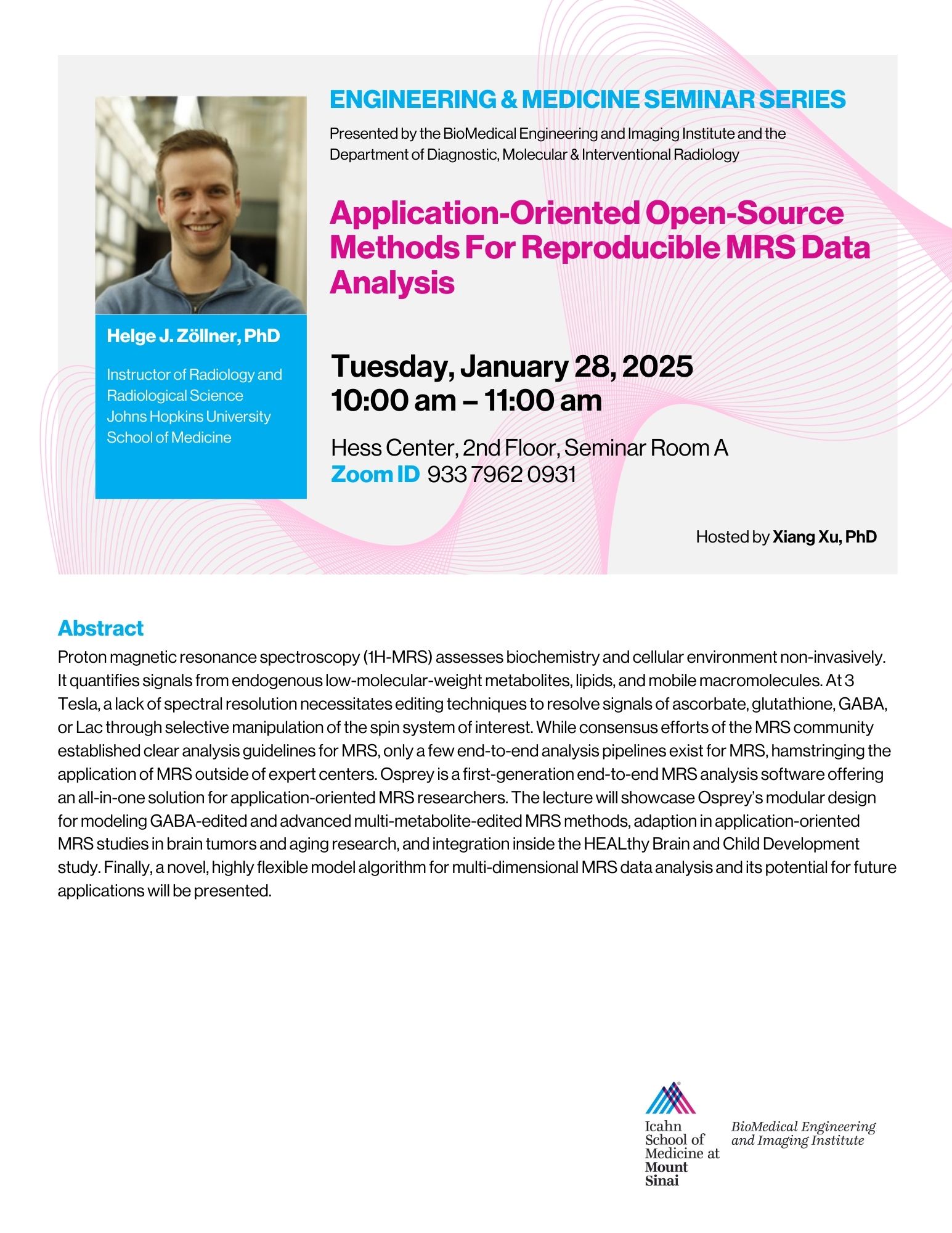
Application-Oriented Open-Source Methods For Reproducible MRS Data Analysis" by Helge J. Zöllner, PhD
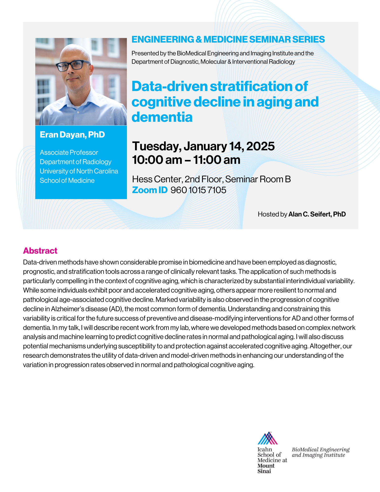
"Data-driven stratification of cognitive decline in aging and dementia" by Eran Dayan, PhD
Data-driven methods have shown considerable promise in biomedicine and have been employed as diagnostic, prognostic, and stratification tools across a range of clinically relevant tasks. The application of such methods is particularly compelling in the context of cognitive aging, which is characterized by substantial interindividual variability. While some individuals exhibit poor and accelerated cognitive aging, others appear more resilient to normal and pathological age-associated cognitive decline. Marked variability is also observed in the progression of cognitive decline in Alzheimer’s disease (AD), the most common form of dementia. Understanding and constraining this variability is critical for the future success of preventive and disease-modifying interventions for AD and other forms of dementia. In my talk, I will describe recent work from my lab, where we developed methods based on complex network analysis and machine learning to predict cognitive decline rates in normal and pathological aging. I will also discuss potential mechanisms underlying susceptibility to and protection against accelerated cognitive aging. Altogether, our research demonstrates the utility of data-driven and model-driven methods in enhancing our understanding of the variation in progression rates observed in normal and pathological cognitive aging.
"Current status of molecular imaging of neuroinflammation: from biomarker to application" by Aisling Chaney, PhD
Abstract
Chronic inflammation and immune dysfunction have emerged as key factors in the pathogenesis of neurological disorders such as Alzheimer’s disease (AD). Non-invasive molecular imaging has the potential to reveal valuable insights into the complex neuroimmune interactions associated with neurodegeneration and injury. Here, we will summarize current and emerging biomarkers for positron emission tomography (PET) imaging of inflammation from cellular and functional specificity to radiotracer development and application.
Bio
Dr. Chaney is an assistant professor at Mallinckrodt Institute of Radiology (MIR) at Washington University in St. Louis. Her research is focused on the development and translation of novel non-invasive molecular imaging strategies to elucidate the inflammatory component of devastating neurological diseases. In particular, she is interested in the relationship between peripheral and central nervous system innate immune responses, and how this crosstalk affects disease development and progression. Dr. Chaney previously worked at Stanford University as a postdoctoral fellow and instructor in the radiology department. She earned her doctorate in neurosciences from the University of Manchester in the United Kingdom.
"Visualization of Histone Deacetylases in the Human Brain" by Changning Wang, PhD
"Magnetic susceptibility MRI for imaging brain iron, myelin and microstructures during neurodegeneration" by Xu Li, PhD
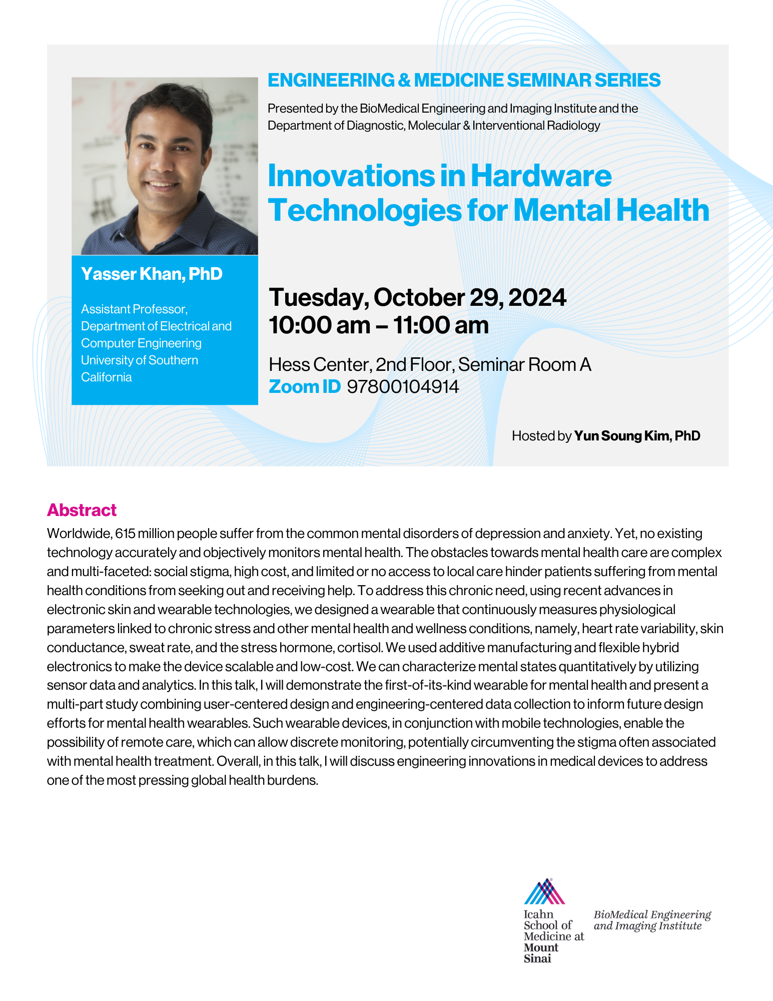
"Innovations in Hardware Technologies for Mental Health" by Yasser Khan, PhD
Please contact us if you would like to access the recording.
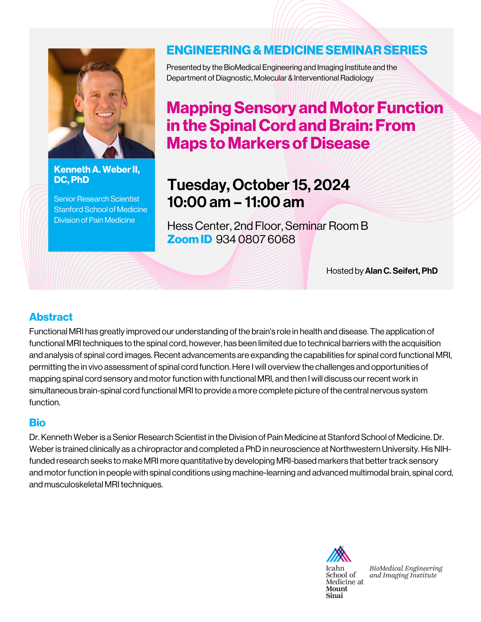
"Mapping Sensory and Motor Function in the Spinal Cord and Brain: From Maps to Markers of Disease" by Kenneth A. Weber II, DC, PhD
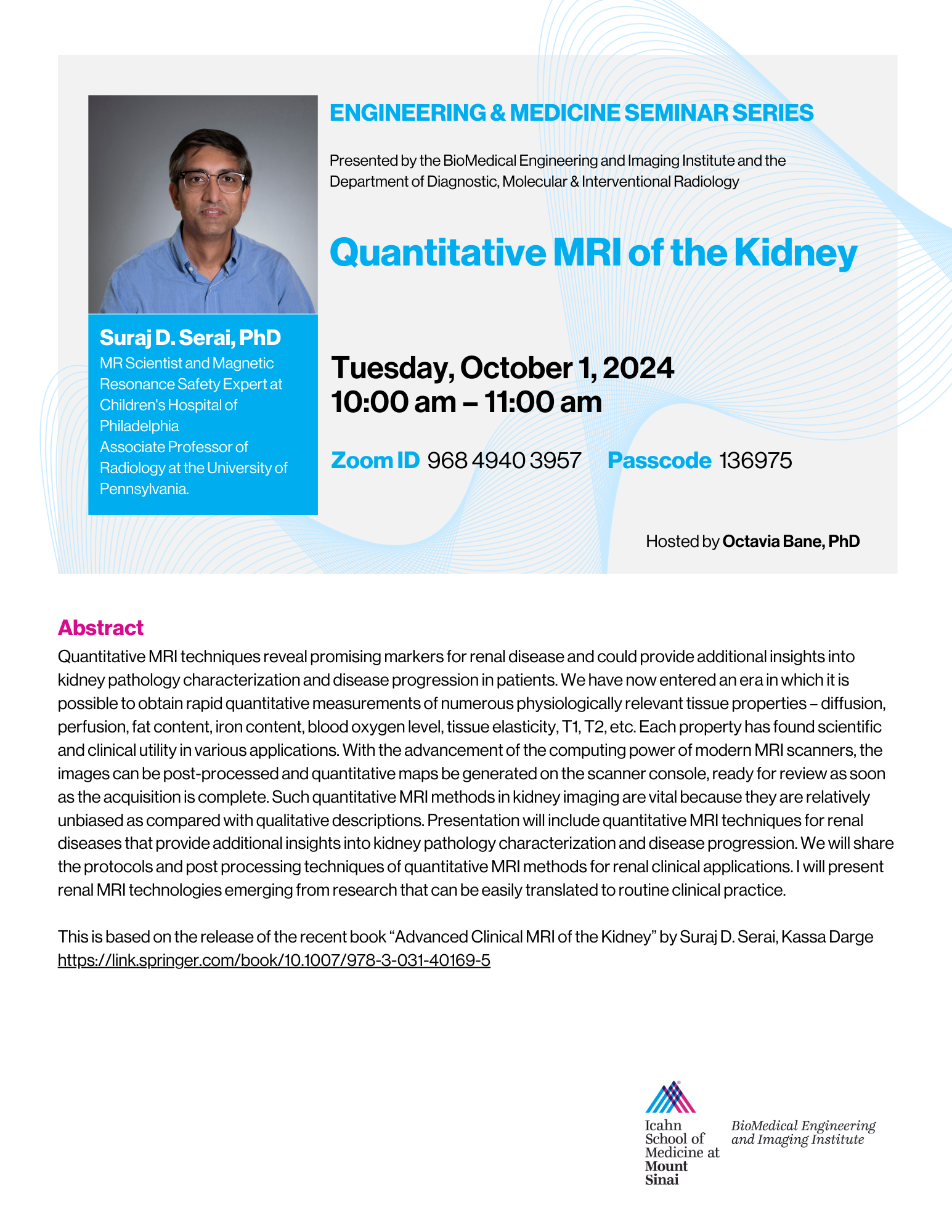
"Quantitative MRI of the Kidney" by Suraj D. Serai, PhD
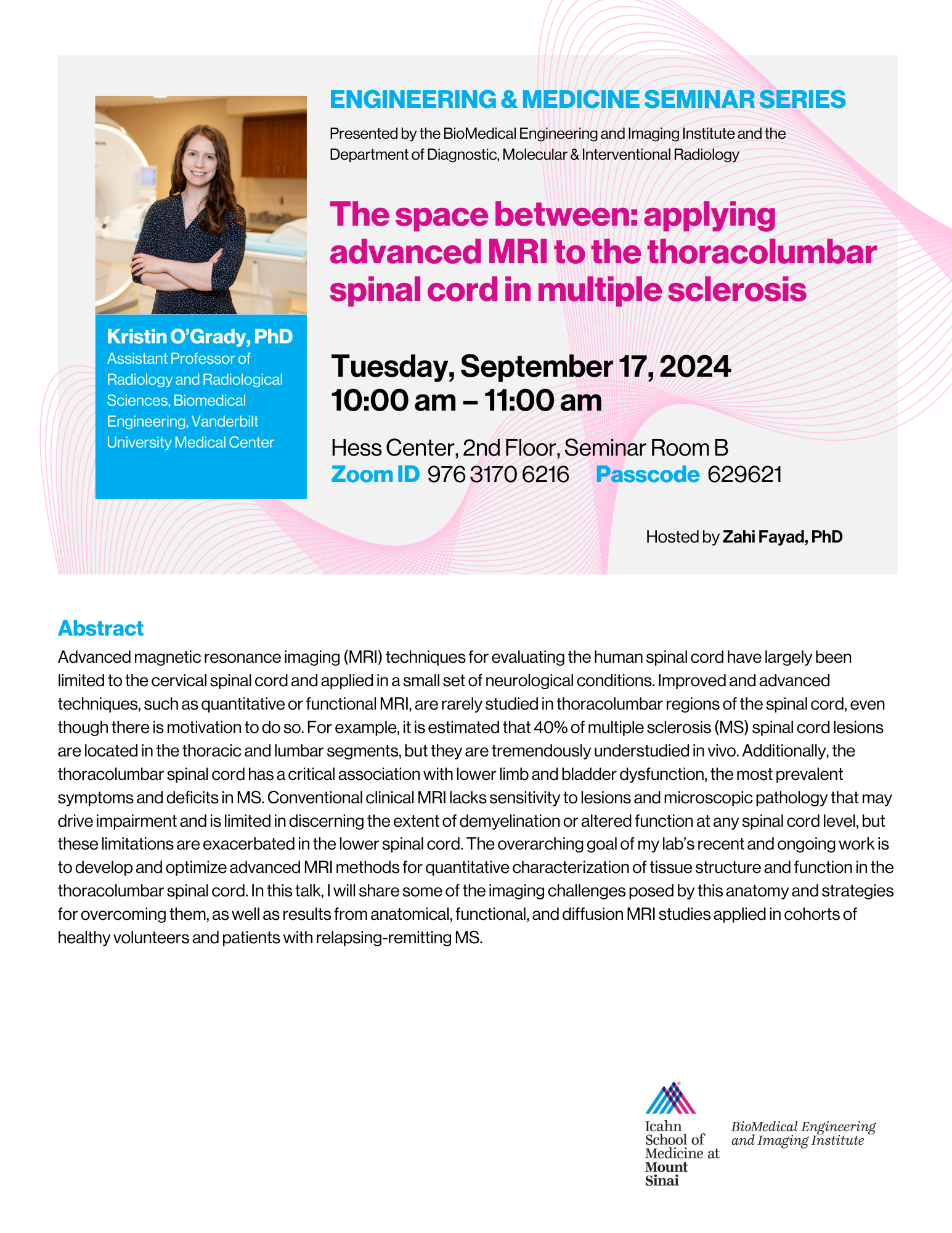
"The space between: applying advanced MRI to the thoracolumbar spinal cord in multiple sclerosis" by Kristin O’Grady, PhD
Computational Medicine for Mental and Physical Health by Rose Faghih, PhD
BMEII Guest Speakers
Below you can see the list of the past BMEII Seminars
"High Resolution Dynamic Imaging: Application to DCE MRI and CEST MRI" by Jaeseok Park, PhD
Abstract
Magnetic resonance imaging has been widely utilised to investigate physiological functions, including functional brain imaging, microvascular dynamics, molecular imaging, etc. In this talk, we introduce rapid high-resolution dynamic MRI exploiting both general spatiotemporal and domain-specific priors for dynamic contrast-enhanced imaging and chemical exchange saturation transfer imaging. Subsequently, we show utilities of these developed techniques on brain cancer and Alzheimer’s Disease.
Bio
Dr. Park is a professor in the Department of Biomedical Engineering at Sungkyunkwan University in the Republic of Korea, where he leads research at the intersection of MR imaging physics, signal processing, and machine learning for brain and cardiac applications. He currently serves as Director of the Brain Korea 21 Project on Intelligent Precision Healthcare Convergence.
Dr. Park brings extensive academic and industry experience, having previously held faculty positions at Korea University and Yonsei University, as well as a research role at Siemens Medical Solutions in Germany. He is Editor-in-Chief of Investigative MRI and serves on the editorial board of the Korean Journal of Radiology. A long-standing member of ISMRM and KSMRM, he has contributed as an ad hoc reviewer for numerous leading journals in the field of medical imaging.
He earned his Ph.D. in Biomedical Engineering from Northwestern University under the mentorship of Dr. Debiao Li.
Recording not available.
"Magnetic Resonance Metabolomics: From Intact Tissues to Imaging and Live-Cell Dynamics" by Leo L. Cheng, PhD
Guided by findings after our discovery of high-resolution magic angle spinning (HRMAS) nuclear magnetic resonance (NMR) spectroscopy, which permits intact analysis of biological tissue and biofluids, our laboratory developed NMR metabolomics. These methodologies can analyze specimens as small as ~1mg for intact tissues, or ~10ul for biofluids (blood, cerebrospinal fluid). We further developed NMR metabolomics, as well as metabolomic imaging, to study malignancy, neurodegeneration, kidney disease, and other illnesses for which diagnosis, staging, and prognostication pose significant clinical challenges. In prostate and lung cancer studies, we showed metabolomics ability to provide important, additional biological parameters to support clinical decision-making. Most recently, our lab demonstrated that our HRMAS spectroscopy methodology can furnish a reaction chamber for monitoring live-cell, real-time dynamic processes of metabolomic reactions, with real-time monitoring of <100,000 cells over 48 hours. Our discussion will highlight metabolomic findings in human lung and prostate cancers, as well as Clostridioides difficile cell real-time dynamics.
"Diffusion Neuroimaging" by Santiago Coelho, PhD
"Evaluating MRI pH mapping as a tool for kidney and cancer imaging" by Michael T. McMahon, PhD
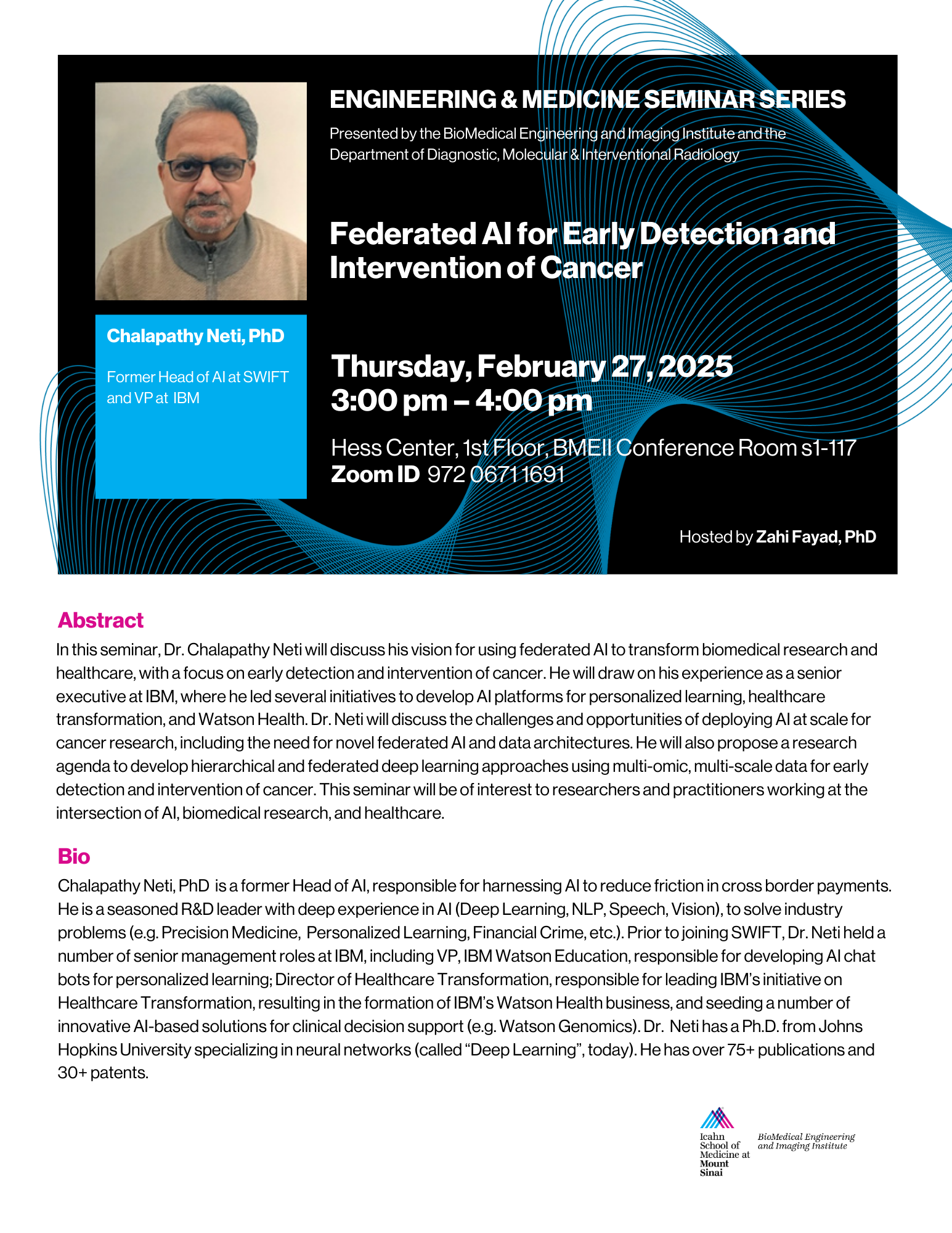
"Federated AI for Early Detection and Intervention of Cancer" by Chalapathy Neti, PhD
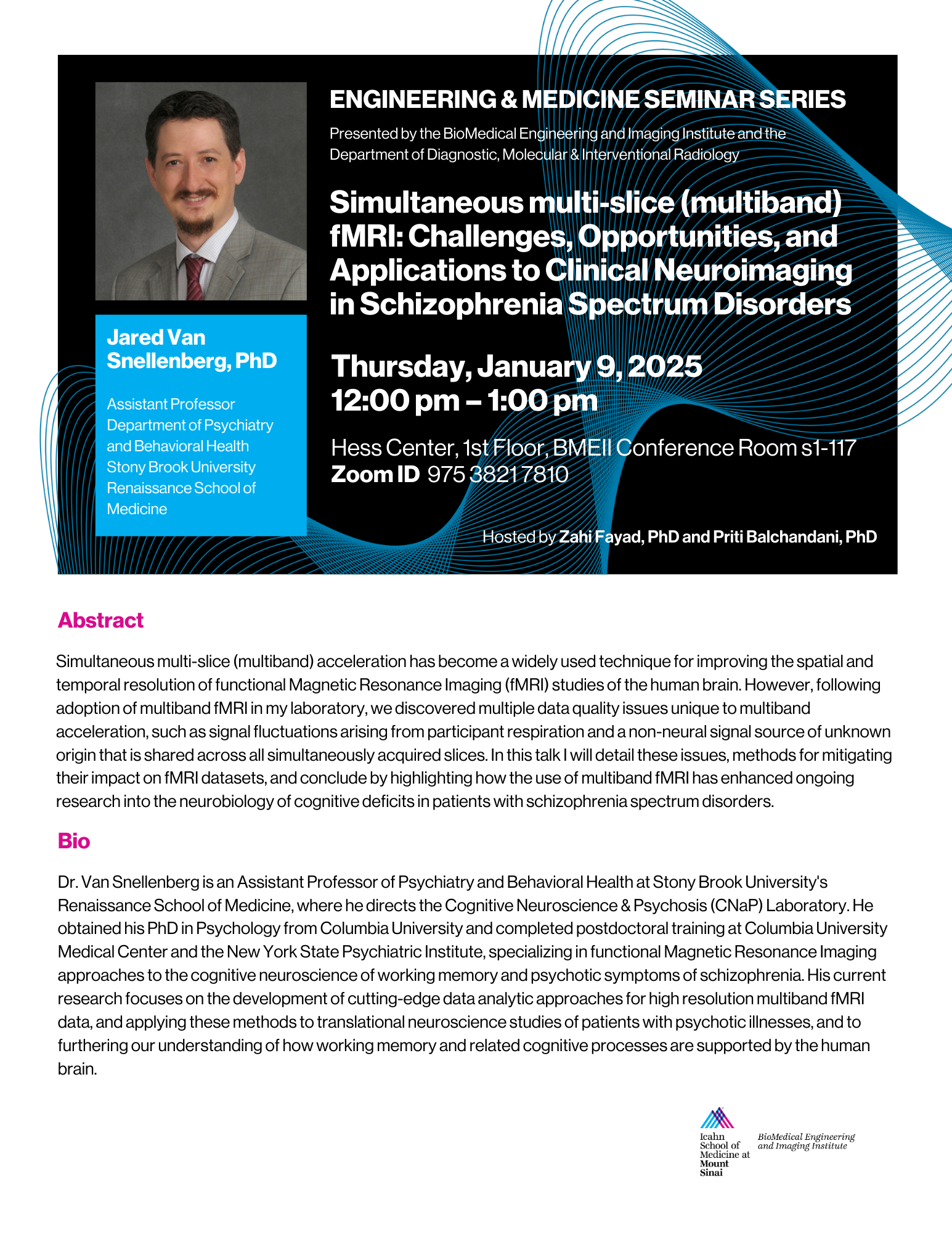
"Simultaneous multi-slice (multiband) fMRI: Challenges, Opportunities, and Applications to Clinical Neuroimaging in Schizophrenia Spectrum Disorders" by Jared Van Snellenberg, PhD
"A Human Cardiovascular Micro-Organ System for Modelling Physiology, Development, and Disease" by David Sachs, PhD
Abstract
Modeling human physiology, development, and disease in vitro is currently accomplished with iPSC derived spheroids and organ-on-chip devices. However, these are limited in that organ-on-chip systems lack important cellular diversity and microphysiology, and spheroids do not capture the tube-like geometry that is essential for most organ functions and multi-organ connectivity. To address the need for more accurate human organ models, we are developing a micro-organ-on-chip system in which iPSCs are seeded into a microfluidic chip and differentiated into tube-shaped organoids in situ, in a system that is guided, but not restricted, by the microfluidic geometry. Starting with the cardiovascular system as a proof of concept, this platform has the potential to simultaneously capture cellular diversity, microphysiology, organ function, and multi-organ connectivity. Custom robotic systems were also developed to control both the seeding and monitoring processes, achieving a controllable variety of micro-organ geometries. As natural organ development leverages continuous feedback from neighboring organ systems, our current engineering efforts are to shift our automation method to deep reinforcement learning, in order to increase the repeatability and throughput of the system via real-time feedback control of organ differentiation.
Bio
David Sachs, PhD, is an Assistant Professor of Genetics and Genomic Sciences at the Icahn School of Medicine at Mount Sinai. He is developing a new micro-organ model of the human cardiovascular system, grown from stem cells inside a microfluidic chip, under the control of a custom robotic platform. The chip will be used to study heart disease and development, including cardiovascular stiffening diseases due to microgravity, and is currently scheduled to launch to the International Space Station in March of 2025. Recent funding will expand the chip with the addition of a liver micro-organ, moving toward the longer term goal of AI guided manufacturing of a comprehensive human micro-organ-on-chip system designed to be readily accessible to a diversity of collaborators. Prior to his work at Mount Sinai, he led the Advanced Application Development group at the MEMS semiconductor startup InvenSense, including the algorithm and digital architecture design of motion sensing devices that have been distributed in billions of units. He has undergraduate degrees in physics and piano performance, a master’s degree from the MIT Media Lab, and a PhD in Biomedical Sciences from Mount Sinai.
Passcode: uv9+0k2y
Lucy G. Moses Lecture
Join us for our 11th Annual Lucy G. Moses Lecture