Neuroimaging
At the BioMedical Engineering and Imaging Institute (BMEII), one of our main areas of research is the development of novel, transformative tools to better visualize and quantify the structure, function and metabolism of the brain. These tools are immediately translated to study the living human brain in the diseased and healthy state.
Over the last three decades, there has been unprecedented progress in the study of the brain. Mount Sinai is one of the few institutions that invest in 7 Tesla (7T) human MRI scanners, which produce high-resolution images to visualize and measure previously undetectable changes in the brain. This advanced imaging technology noninvasively captures subtle neurological abnormalities, enabling early detection of disease and potential treatments for patients with illnesses that are resistant to medication.
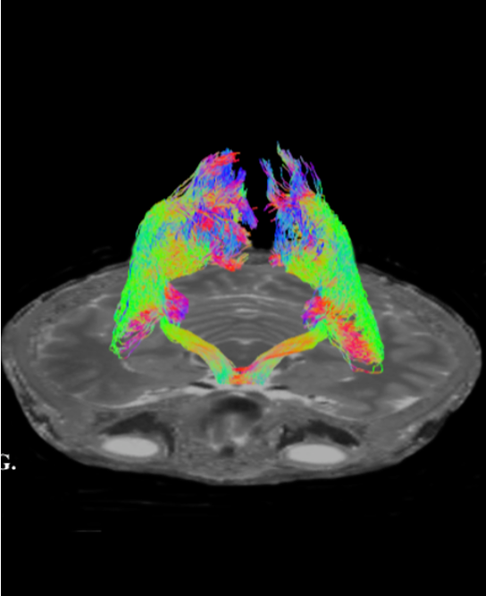
Our research improves the performance of all neuroimaging modalities. This includes functional MRI, spinal cord imaging, diffusion MRI to study brain connectivity and perform tractography, and MR spectroscopic imaging for the study of metabolism.
Creative engineering solutions in imaging transform the management of neurological diseases with complex or subtle pathology, expand the use of minimally invasive treatment options, and bring us one step closer to unraveling the mysteries of the human brain.
Brain imaging research is overseen by the Advanced Neuroimaging Research Program (ANRP), led by Dr. Priti Balchandani, Associate Professor of Radiology and Neuroscience. ANRP focuses on developing novel imaging technologies to diagnose and treat a wide range of conditions, including epilepsy, brain tumors, psychiatric illnesses, multiple sclerosis, and spinal cord injury.
Leadership
Priti Balchandani, PhD, is a Professor of Diagnostic, Molecular and Interventional Radiology, Neuroscience and Psychiatry. She is the Director of the Advanced Neuroimaging Research Program (ANRP) and Associate Director of the BioMedical Engineering and Imaging Institute (BMEII). Her research has been focused on bridging the gap between advanced electrical engineering techniques and medical imaging applications.

Photo Credit: Claudia Paul
Advanced Neuroimaging
Research Program
Leveraging technical and clinical advantages of
ISMMS to facilitate the most innovative
brain imaging research
Learn More
Ultrahigh Field MRI Group
Devising creative engineering methods to overcome
key limitations of operating at ultra high magnetic fields,
capitalize on signal, and contrast advantage to visualize
the human brain in exceptional detail
Learn More
Chemical Exchange Saturation Transfer Imaging
Enabling the detection of specific metabolites indirectly through water via the exchange process
Click for more information
Glucose uptake is dynamically monitored, through careful design and optimization, by MRI. Studying tumor regions in mice implanted with human brain tumors and injected with glucose, signal enhancements are observed. This glucose imaging technique is also used in the study of perfusion, permeability properties in animal models, and humans at 7T and 3T during an intravenous glucose tolerance test. Our research, led by Dr. Xiang Xu, aims to utilize this contrast mechanism to study the integrity of the blood brain barrier in brain tumors and neurodegenerative diseases, such as multiple sclerosis.
Multiscale Imaging From In Vivo
to Ex Vivo and Brain, Brainstem,
and Spinal Cord 7T MRI
Extending 7-Tesla MRI to the brainstem and spinal cord,
and exploring ultrahigh resolution imaging of
ex vivo brain samples
Click for more information
Dr. Alan Seifert, a biomedical engineer with a background in both imaging physics and the medical sciences, conducts essential research related to in vivo and ex vivo imaging and 7T MRI. The central theme of his research is to identify biologically relevant magnetic resonance imaging (MRI) signals that are particularly challenging to detect, and develop imaging techniques and hardware that can quantify these signals, thus providing new and clinically valuable imaging biomarkers.
His current work is focused in three main areas, all related to advancing our understanding of the relationship between the structure and composition of neural tissue and the functional integrity of neural circuits. First, he develops anatomical, functional, and diffusion MRI methods to study neural circuits spanning the spinal cord, brainstem, cerebellum, and brain, particularly those involved in pain modulation. He also refines and applies high-resolution multi-contrast and quantitative ex vivo MRI methods to complement and enhance neuropathological research. Finally, he works to establish and promote solid-state MRI methods, such as ultrashort echo time (UTE) and zero echo time (ZTE), for direct quantification of myelin concentration.
Abnormal Brain Pathologies
Using functional and structural high-field 7T MRI
to study the pathology of neurological diseases and
psychiatric disorders
Click for more information
We approach the study of abnormal brain pathologies using advanced 7T MRI technology. Dr. Laurel Morris, Director of the Morris Lab and Assistant Professor in the Department of Psychiatry at ISMMS, develops neurocognitive assessments for in-depth characterizations of brain function during optimized high-field 7T MRI scans.
Dr. Yael Jacob, Assistant Professor in the Department of Psychiatry, researches the development and implementation of new computational methods for the study of complex brain networks using functional and structural MRI. She specializes in the mathematical field of graph theory, applying it to the study of interaction between neural networks during different cognitive tasks and their differing manifestation among brain pathologies. These methods are used to decipher brain network mechanisms that underlie behavioral and pathological differences in different neurological diseases and psychiatric disorders.
Faculty - Biomedical Engineering and Regenerative Medicine
The BioMedical Engineering and Imaging Institute (BMEII) at the Icahn School of Medicine at Mount Sinai (ISMMS), in partnership with Rensselaer Polytechnic Institute, is seeking mid to senior-level faculty members to spearhead a novel research initiative in the engineering of reparative and regenerative Medicine.
Faculty - Artificial Intelligence (AI) and Translation to Medicine
Faculty - Neuroengineering
Postdoctoral Fellow
The BioMedical Engineering and Imaging Institute at the Icahn School of Medicine at Mount Sinai is seeking a Postdoctoral Fellow.
News
Mount Sinai Researchers Featured in Nature for Advancing Healthspan Science
Nature has published a sponsored feature highlighting Mount Sinai’s leadership in redefining ageing through the XPRIZE Healthspan initiative. The article showcases the work of Drs. Miriam Merad, Zahi Fayad, and Fanny Elahi, who are pioneering new approaches to extend...
Dr. Zahi Fayad Featured in TIME for Innovative Research on Health Tracking and Longevity (Digital Twin Study)
“Let’s say you are stable, stable, stable, then suddenly start seeing a small dip in some measurements. That dip is presymptomatic, by the way ... That’s when I want to intervene.” — Zahi Fayad
[VIDEO] Dr. Zahi Fayad at Aspen Ideas 2025: Health Panel on Healthspan
What is health—and how do we measure it? Dr. Zahi Fayad emphasizes extending healthspan, which focuses on optimizing human health across the lifespan. By defining and tracking the trajectory of health, we can intervene earlier, and ensure people live not just longer,...
Latest Publications
Artificial intelligence–enabled rapid diagnosis of patients with COVID-19
Xueyan Mei, Hao-Chih Lee, […] Yang Yang
Nat Med (2020).
Probing myeloid cell dynamics in ischaemic heart disease by nanotracer hot-spot imaging
Max L. Senders, Anu E. Meerwaldt, ... Willem J. M. Mulder
Nat. Nanotechnol. 15, 398–405 (2020).