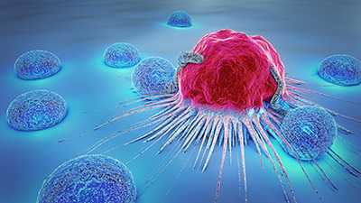
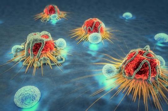
Cancer Imaging
Researchers at the BMEII Imaging Research Center are currently undertaking groundbreaking studies in the use of imaging for the early detection and treatment assessment of cancer. Early results validate the use of noninvasive imaging tools for the detection of and screening for cancer. BMEII staff use full-body MRI service that measures body waves and blood flow to develop mathematical and statistical models to determine characteristics of a variety of cancers.
Our specialists at BMEII are studying how MRI signals affect oxygen in blood and tumor tissue. Our group images human subject in a newly opened research facility with the state-of-the-art 3 Tesla MRI system.
The Cancer Imaging Core at BMEII aims to develop, organize, and provide imaging support for cancer imaging trials in collaboration with Tisch Cancer Center oncologists. This includes providing protocol expertise, scheduling imaging studies, and assessing tumor response in a timely manner. Tumor assessment is now provided electronically using MINT software. New imaging methods are being developed that will allow clinicians not only to see where a tumor is located in the body, but also to visualize the expression and activity of specific molecules that influence tumor behavior and/or response to therapy. This information is expected to have a major impact on cancer detection, individualized treatment, and drug development, as well as on our understanding of how cancer arises. We intend to narrow or close the gap between science and patients.
Core Members:
• Bachir Taouli, MD, MHA – Professor of Diagnostic, Molecular and Interventional Radiology and Medicine/Vice-chair of Translational Research
• Jordan Cuevas – Clinical Research Manager
Methods currently used within the Cancer Core:
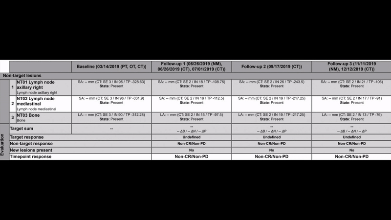
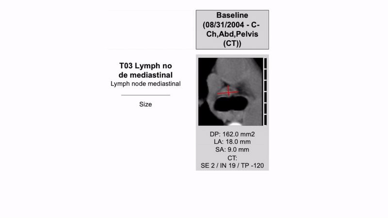
RECIST – A standard way to measure the response of a tumor to treatment. If a study is eligible, one must choose target lesions that are easy to measure and calculate the sum of the length diameter.
Below is an example of a RECIST’s exam findings
CHESON – The evaluation of Lymphoma which Includes clinical examination, BM biopsy, CT scan and PET scan for lymphomas that are frequently hyper metabolic.
Below is an example of a Cheson Reading
News
[VIDEO] Aspen Ideas Festival: Dr. Fuchs and Dr. Fayad on Powering Medicine with Technology and Data Science
Zahi A. Fayad, PhD, Director of Biomedical Engineering and Imaging Institute sits down with Thomas Fuchs, DrSc, Dean of Artificial Intelligence and Human Health to discuss powering medicine with technology and data science. Click the image to watch the video.
[VIDEO] Dr. Zahi Fayad: Powering Medicine with Technology and Data Science
BMEII Director Zahi Fayad discusses the role of data science in medicine at the 2022 Aspen Ideas Festival. Click the image to watch the video.
Latest Publications
Artificial intelligence–enabled rapid diagnosis of patients with COVID-19
Xueyan Mei, Hao-Chih Lee, […] Yang Yang
Nat Med (2020).
Probing myeloid cell dynamics in ischaemic heart disease by nanotracer hot-spot imaging
Max L. Senders, Anu E. Meerwaldt, ... Willem J. M. Mulder
Nat. Nanotechnol. 15, 398–405 (2020).